2
views
0
recommends
+1 Recommend
0 collections
    0
    shares
      • Record: found
      • Abstract: found
      • Article: found
      Is Open Access

      Assessing small airway disease in GLI versus NHANES III based spirometry using area under the expiratory flow-volume curve

      research-article

      Read this article at

      Bookmark
          There is no author summary for this article yet. Authors can add summaries to their articles on ScienceOpen to make them more accessible to a non-specialist audience.

          Abstract

          Background

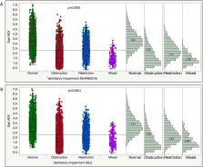
          Spirometry interpretation is influenced by the predictive equations defining lower limit of normal (LLN), while ‘distal’ expiratory flows such as forced expiratory flow at 50% FVC (FEF 50) are important functional parameters for diagnosing small airway disease (SAD). Area under expiratory flow-volume curve (AEX) or its approximations have been proposed as supplemental spirometric assessment tools. We compare here the performance of AEX in differentiating between normal, obstruction, restriction, mixed defects and SAD, as defined by Global Lung Initiative (GLI) or National Health and Nutrition Examination Survey (NHANES) III reference values, and using various predictive equations for FEF 50.

          Methods

          We analysed 15 308 spirometry-lung volume tests. Using GLI versus NHANES III LLNs, and diagnosing SAD by the eight most common equation sets for forced expiratory flow at 50% of vital capacity lower limits of normal (FEF 50 LLN), we assessed the degree of diagnostic concordance and the ability of AEX to differentiate between various definition-dependent patterns.

          Results

          Concordance rates between NHANES III and GLI-based classifications were 93.7%, 78.6%, 86.8%, 88.0%, 93.8% and 98.8% in those without, with mild, moderate, moderately severe, severe and very severe obstruction, respectively (agreement coefficient 0.81 (0.80–0.82)). The prevalence of SAD was 0.6%–6.9% of the cohort, depending on the definition used. The AEX differentiated well between normal, obstruction, restriction, mixed pattern and SAD, as defined by most equations.

          Conclusions

          If the SAD diagnosis is established by using mean FEF 50 LLN or a set number of predictive equations, AEX is able to differentiate well between various spirometric patterns. Using the most common predictive equations (NHANES III and GLI), the diagnostic concordance for functional type and obstruction severity is high.

          Related collections

          Most cited references25

          • Record: found
          • Abstract: not found
          • Article: not found

          Lung function testing: selection of reference values and interpretative strategies. American Thoracic Society.

            Bookmark
            • Record: found
            • Abstract: found
            • Article: not found

            Impulse oscillometry: interpretation and practical applications.

            Simple spirometry and body plethysmography have been routinely used in children aged > 5 years. New techniques based on physiologic concepts that were first described almost 50 years ago are emerging in research and in clinical practice for measuring pulmonary function in children. These techniques have led to an increased understanding of the pediatric lung and respiratory mechanics. Impulse oscillometry (IOS), a simple, noninvasive method using the forced oscillation technique, requires minimal patient cooperation and is suitable for use in both children and adults. This method can be used to assess obstruction in the large and small peripheral airways and has been used to measure bronchodilator response and bronchoprovocation testing. New data suggest that IOS may be useful in predicting loss of asthma control in the pediatric population. This article reviews the clinical applications of IOS, with an emphasis on the pediatric setting, and discusses appropriate coding practices for the clinician.
              Bookmark
              • Record: found
              • Abstract: found
              • Article: not found

              Lung volumes in healthy nonsmoking adults.

              Total lung capacity (TLC), functional residual capacity, residual volume, and corresponding 95% confidence intervals were measured in 245 healthy nonsmoking person (122 women, 123 men) using a single-breath helium technique. Prediction equations for lung volumes were generated by multiple linear regression. The resultant equations are similar to previously published equations using multiple-breath gas equilibration techniques. Measured 95% confidence intervals can be closely approximated by using two times the standard error of the estimate for each equation, but cannot be approximated by using +/- 20% of the predicted value. Radiographic TLC was not significantly different from the helium dilution TLC.
                Bookmark

                Author and article information

                Journal
                BMJ Open Respir Res
                BMJ Open Respir Res
                bmjresp
                bmjopenrespres
                BMJ Open Respiratory Research
                BMJ Publishing Group (BMA House, Tavistock Square, London, WC1H 9JR )
                2052-4439
                2019
                24 November 2019
                : 6
                : 1
                : e000511
                Affiliations
                [1 ]departmentDivision of Pulmonary, Allergy, Critical Care and Sleep Medicine , School of Medicine, Emory University , Atlanta, Georgia, USA
                [2 ]Section of Sleep Medicine, Atlanta VAMC , Atlanta, Georgia, USA
                [3 ]Respiratory Institute, Cleveland Clinic , Cleveland, Ohio, USA
                Author notes
                [Correspondence to ] Dr Octavian C Ioachimescu; oioac@ 123456yahoo.com
                Author information
                http://orcid.org/0000-0001-9047-6894
                Article
                bmjresp-2019-000511
                10.1136/bmjresp-2019-000511
                6890381
                a5ef0773-ebb4-487d-ad40-e251416d76c3
                © Author(s) (or their employer(s)) 2019. Re-use permitted under CC BY-NC. No commercial re-use. See rights and permissions. Published by BMJ.

                This is an open access article distributed in accordance with the Creative Commons Attribution Non Commercial (CC BY-NC 4.0) license, which permits others to distribute, remix, adapt, build upon this work non-commercially, and license their derivative works on different terms, provided the original work is properly cited, appropriate credit is given, any changes made indicated, and the use is non-commercial. See:  http://creativecommons.org/licenses/by-nc/4.0/.

                History
                : 07 October 2019
                : 31 October 2019
                : 01 November 2019
                Categories
                Respiratory Physiology
                1506
                2230
                Custom metadata
                unlocked

                lung physiology,respiratory measurement,asthma,lung transplantation

                Comments

                Comment on this article