27
views
0
recommends
+1 Recommend
0 collections
    0
    shares
      • Record: found
      • Abstract: found
      • Article: found
      Is Open Access

      Primary Angiosarcoma of the Spleen: An Oncological Enigma

      case-report

      Read this article at

      Bookmark
          There is no author summary for this article yet. Authors can add summaries to their articles on ScienceOpen to make them more accessible to a non-specialist audience.

          Abstract

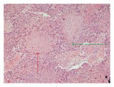
          Introduction. Primary splenic angiosarcoma is an extremely unusual neoplasm originating from sinusoidal vascular endothelium. Surgical extirpation is the mainstay of treatment of this highly malignant disease. Case Presentation. An 82-year-old woman was admitted with left pleural effusion and a palpable left upper quadrant abdominal mass, secondary to splenomegaly by two large splenic tumors. Classic open splenectomy was performed and angiosarcoma of the spleen was the final histopathological diagnosis, which was primary since no other disease site was revealed. Discussion. The incidence of the disease is 0.14–0.23 cases per million, with slight male predominance. Etiology is not established and clinical presentation may confuse even experienced physicians. Imaging modalities cannot differentiate the lesion from other vascular splenic neoplasms and the correct diagnosis is mainly set after histopathological examination of the resected spleen. As with other sarcomas, surgery is the only curative approach, while chemo- and radiotherapy have poor results. Prognosis remains dismal.

          Related collections

          Most cited references24

          • Record: found
          • Abstract: found
          • Article: not found

          Angiosarcoma. A report of 67 patients and a review of the literature.

          Angiosarcomas (AS) are rare, aggressive tumors. Optimal treatment has not been well defined. The authors undertook a retrospective review of patients seen at their institution with the intent of identifying prognostic factors and optimal treatment strategies. Between 1955 and 1990, 67 patients with AS were seen at the University of California, at Los Angeles Medical Center. Follow-up ranged from 1 to 173 months with a median of 30 months. The overall prognosis was poor. The actuarial 2- and 5-year disease free survivals (DFS) were 44% and 24%, respectively. Of 52 recurrences after primary treatment, 81% (42 of 52) had a component of local failure. Twenty-eight patients had developed distant metastases at last follow-up. Of patients who received surgery (S) and radiation therapy (RT), with or without chemotherapy (CT), 5-year actuarial DFS was 43%, compared with 17% for patients who underwent S +/- CT as initial treatment (P = 0.03). Only 9% of patients (1 of 11) treated with RT +/- CT were rendered free of disease. Patients with AS usually present with high grade histology, and with multifocal disease. There is a propensity for both local recurrence and distant metastases. Our results and a review of the literature, suggest that S plus RT offers the best chance for long term control of this aggressive tumor. The role of CT remains undefined.
            Bookmark
            • Record: found
            • Abstract: found
            • Article: not found

            Primary angiosarcoma of the spleen. A clinicopathologic study of 40 cases.

            Forty primary splenic angiosarcomas occurring in 21 men and 19 women, 19-84 years old (median 59 years) are reported. Patients presented with splenomegaly (35 of 38, 92%), abdominal pain (33 of 40, 83%), and systemic symptoms such as fatigue (2 of 40, 5%), fever (4 of 40, 10%), and/or weight loss (16 of 40, 40%). Five (13%) experienced splenic rupture associated with hemoperitoneum. Abnormal laboratory findings included cytopenia (31 of 34, 91%), leukocytosis (8 of 21, 38%), and thrombocytosis (1/39, 3%). Most spleens weighed 500-1,000 g (mean, 1,180 g). The cut splenic surfaces showed multiple hemorrhagic nodules that were frequently associated with infarction, although some had a diffuse pattern of involvement. Microscopically, there were a variety of histologic patterns displayed by the vasoformative component. A honeycomb or sponge-like pattern was common in some, whereas others simulated a cavernous hemangioma or normal splenic sinuses (pseudosinusoidal pattern). Papillary endothelial tufts and solid proliferations of spindled to round to epithelioid cells were also seen. Factor VIII-related antigen was detected in 19 of 23 cases, BMA-120 in 18 of 23, UEA-1 receptor in 18 of 23, and vimentin in 23 of 23 as well as CD68 antigen in 1 of 23 cases. S-100 protein and cytokeratin were not found in any of the 23 cases studied. Metastases in 22 of 32 patients (69%) were to the liver (13 patients), bone or bone marrow (7 patients), lymph nodes (1 patient), and brain (1 patient). Three patients had concomitant malignancies and one had a prior history of a mixed B-cell lymphoma 5 years previously that had been treated with chemotherapy. Follow-up in 38 patients revealed that 30 (79%) are dead at a median interval of 6 months (range 0-48 months) and 8 are alive 5-21 months after diagnosis. These findings indicate that splenic angiosarcoma is an aggressive neoplasm with a high metastatic rate and an abysmal prognosis. Recognition of the wide range of histologic patterns is of diagnostic value but no apparent prognostic significance.
              Bookmark
              • Record: found
              • Abstract: found
              • Article: not found

              Splenic angiosarcoma: a clinicopathologic and immunophenotypic study of 28 cases.

              Primary angiosarcoma of the spleen is a rare neoplasm that has not been well characterized. We describe the clinical, morphologic, and immunophenotypic findings of 28 cases of primary splenic angiosarcoma, including one case that shares features of lymphangioma/lymphangiosarcoma. The patients included 16 men and 12 women, aged 29 to 85 years, with a mean of 59 years and median of 63 years. The majority of patients (75%) complained of abdominal pain, and 25% presented with splenic rupture. The most common physical finding was splenomegaly (71%). Seventeen of 21 patients were reported to have anemia. Macroscopic examination showed splenomegaly in 85% cases. Sectioning revealed discrete lesions in 88% of cases, ranging from well-circumscribed firm nodules to poorly delineated foci of necrosis and hemorrhage associated with cystic spaces. Microscopically, the tumors were heterogenous; however, all cases demonstrated at least a focal vasoformative component lined by atypical endothelial cells. Solid sarcomatous, papillary, and epithelioid growth patterns were observed. The solid sarcomatous component resembled fibrosarcoma in two cases and malignant fibroushistiocytoma in one case. Hemorrhage, necrosis, hemosiderin, extramedullary hematopoiesis, and intracytoplasmic hyaline globules were frequently identified. A panel of immunohistochemical studies revealed that the majority of tumors were immunoreactive for at least two markers of vascular differentiation (CD34, FVIIIRAg, VEGFR3, and CD31) and at least one marker of histiocytic differentiation (CD68 and/or lysozyme). Metastases developed in 100% of patients during the course of their disease. Twenty-six patients died of disease despite aggressive therapy, whereas only two patients are alive at last follow-up, one with disease at 8 years and the other without disease at 10 years. In conclusion, primary splenic angiosarcoma is an extremely aggressive neoplasm that is almost universally fatal. The majority of splenic angiosarcomas coexpress histiocytic and endothelial markers by immunohistochemical analysis, which suggest that some tumors may originate from splenic lining cells.
                Bookmark

                Author and article information

                Journal
                Case Rep Oncol Med
                Case Rep Oncol Med
                CRIONM
                Case Reports in Oncological Medicine
                Hindawi Publishing Corporation
                2090-6706
                2090-6714
                2014
                2 July 2014
                : 2014
                : 193036
                Affiliations
                1Department of Pathology, Tzaneio General Hospital, 185 36 Piraeus, Greece
                22nd Department of Surgery, Aretaieion Hospital, 115 28 Athens, Greece
                32nd Department of Surgery, Tzaneio General Hospital, 185 36 Piraeus, Greece
                Author notes
                *Myoteri Despoina: dmyoteri@ 123456gmail.com

                Academic Editor: Ossama W. Tawfik

                Article
                10.1155/2014/193036
                4101939
                a62686ba-2c14-4a05-b419-e6b0c608f0e9
                Copyright © 2014 Myoteri Despoina et al.

                This is an open access article distributed under the Creative Commons Attribution License, which permits unrestricted use, distribution, and reproduction in any medium, provided the original work is properly cited.

                History
                : 28 March 2014
                : 8 June 2014
                : 15 June 2014
                Categories
                Case Report

                Oncology & Radiotherapy
                Oncology & Radiotherapy

                Comments

                Comment on this article