11
views
0
recommends
+1 Recommend
0 collections
    0
    shares
      • Record: found
      • Abstract: found
      • Article: found
      Is Open Access

      Clinical Feasibility of Synthetic Magnetic Resonance Imaging in the Diagnosis of Internal Derangements of the Knee

      research-article
      , MD, , MD , , MD, , MD
      Korean Journal of Radiology
      The Korean Society of Radiology
      Synthetic MRI, Knee, Cruciate ligament, Meniscus, Cartilage

      Read this article at

      Bookmark
          There is no author summary for this article yet. Authors can add summaries to their articles on ScienceOpen to make them more accessible to a non-specialist audience.

          Abstract

          Objective

          To evaluate the feasibility of synthetic magnetic resonance imaging (MRI) compared to conventional MRI for the diagnosis of internal derangements of the knee at 3T.

          Materials and Methods

          Following Institutional Review Board approval, image sets of conventional and synthetic MRI in 39 patients were included. Two musculoskeletal radiologists compared the image sets and qualitatively analyzed the images. Subjective image quality was assessed using a four-grade scale. Interobserver agreement and intersequence agreement between conventional and synthetic images for cartilage lesions, tears of the cruciate ligament, and tears of the meniscus were independently assessed using Kappa statistics. In patients who underwent arthroscopy (n = 8), the sensitivity, specificity, and accuracy for evaluated internal structures were calculated using arthroscopic findings as the gold standard.

          Results

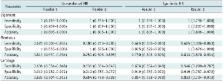
          There was no statistically significant difference in image quality ( p = 0.90). Interobserver agreement (κ = 0.649– 0.981) and intersequence agreement (κ = 0.794–0.938) were nearly perfect for all evaluated structures. The sensitivity, specificity, and accuracy for detecting cartilage lesions (sensitivity, 63.6% vs. 54.6–63.6%; specificity, 91.9% vs. 91.9%; accuracy, 83.3–85.4% vs. 83.3–85.4%) and tears of the cruciate ligament (sensitivity, specificity, accuracy, 100% vs. 100%) and meniscus (sensitivity, 50.0–62.5% vs. 62.5%; specificity, 100% vs. 87.5–100%; accuracy, 83.3–85.4% vs. 83.3–85.4%) were similar between the two MRI methods.

          Conclusion

          Conventional and synthetic MRI showed substantial to almost perfect degree of agreement for the assessment of internal derangement of knee joints. Synthetic MRI may be feasible in the diagnosis of internal derangements of the knee.

          Related collections

          Most cited references32

          • Record: found
          • Abstract: found
          • Article: found
          Is Open Access

          Simultaneous multislice (SMS) imaging techniques

          Simultaneous multislice imaging (SMS) using parallel image reconstruction has rapidly advanced to become a major imaging technique. The primary benefit is an acceleration in data acquisition that is equal to the number of simultaneously excited slices. Unlike in‐plane parallel imaging this can have only a marginal intrinsic signal‐to‐noise ratio penalty, and the full acceleration is attainable at fixed echo time, as is required for many echo planar imaging applications. Furthermore, for some implementations SMS techniques can reduce radiofrequency (RF) power deposition. In this review the current state of the art of SMS imaging is presented. In the Introduction, a historical overview is given of the history of SMS excitation in MRI. The following section on RF pulses gives both the theoretical background and practical application. The section on encoding and reconstruction shows how the collapsed multislice images can be disentangled by means of the transmitter pulse phase, gradient pulses, and most importantly using multichannel receiver coils. The relationship between classic parallel imaging techniques and SMS reconstruction methods is explored. The subsequent section describes the practical implementation, including the acquisition of reference data, and slice cross‐talk. Published applications of SMS imaging are then reviewed, and the article concludes with an outlook and perspective of SMS imaging. Magn Reson Med 75:63–81, 2016. © 2015 The Authors. Magnetic Resonance in Medicine Published by Wiley Periodicals, Inc. on behalf of International Society of Medicine in Resonance.
            Bookmark
            • Record: found
            • Abstract: found
            • Article: not found

            Motion correction with PROPELLER MRI: application to head motion and free-breathing cardiac imaging.

            James Pipe (1999)
            A method for motion correction, involving both data collection and reconstruction, is presented. The PROPELLER MRI method collects data in concentric rectangular strips rotated about the k-space origin. The central region of k-space is sampled for every strip, which (a) allows one to correct spatial inconsistencies in position, rotation, and phase between strips, (b) allows one to reject data based on a correlation measure indicating through-plane motion, and (c) further decreases motion artifacts through an averaging effect for low spatial frequencies. Results are shown in which PROPELLER MRI is used to correct for bulk motion in head images and respiratory motion in nongated cardiac images. Magn Reson Med 42:963-969, 1999. Copyright 1999 Wiley-Liss, Inc.
              Bookmark
              • Record: found
              • Abstract: found
              • Article: not found

              MR imaging of the menisci and cruciate ligaments: a systematic review.

              To systematically review and synthesize published data on the diagnostic performance of magnetic resonance (MR) imaging of the menisci and cruciate ligaments and to assess the effect of study design characteristics and magnetic field strength on diagnostic performance. Articles published between 1991 and 2000 were included if at least 30 patients were studied, arthroscopy was the reference standard, the magnetic field strength was reported, positivity criteria were defined, and the absolute numbers of true-positive, false-negative, true-negative, and false-positive results were available or derivable. Pooled weighted and summary receiver operating characteristic (ROC) analyses were performed for tears of both menisci and both cruciate ligaments separately and for the four lesions combined, by using random effects models. Differences were assessed according to lesion type. Twenty-nine of 120 retrieved articles were included. Pooled weighted sensitivity was higher for medial meniscal tears than that for lateral meniscal tears. However, pooled weighted specificity for the medial meniscus was lower than that for the lateral meniscus. In summary ROC analyses performed per lesion, various study design characteristics were found to influence diagnostic performance. Higher magnetic field strength significantly improved discriminatory power only for anterior cruciate ligament tears. When all lesions were combined in one overall summary ROC analysis, magnetic field strength was a significant but modest predictor of diagnostic performance. Diagnostic performance of MR imaging of the knee is different according to lesion type and is influenced by various study design characteristics. Higher magnetic field strength modestly improves diagnostic performance, but a significant effect was demonstrated only for anterior cruciate ligament tears.
                Bookmark

                Author and article information

                Journal
                Korean J Radiol
                Korean J Radiol
                KJR
                Korean Journal of Radiology
                The Korean Society of Radiology
                1229-6929
                2005-8330
                Mar-Apr 2018
                22 February 2018
                : 19
                : 2
                : 311-319
                Affiliations
                Department of Radiology, Research Institute of Radiological Science, YUHS-KRIBB Medical Convergence Research Institute, and Severance Biomedical Science Institute, Yonsei University College of Medicine, Seoul 03722, Korea.
                Author notes
                Corresponding author: Young Han Lee, MD, Department of Radiology, Research Institute of Radiological Science, YUHS-KRIBB Medical Convergence Research Institute, and Severance Biomedical Science Institute, Yonsei University College of Medicine, 50-1 Yonsei-ro, Seodaemun-gu, Seoul 03722, Korea. Tel: (822) 2228-7420, Fax: (822) 393-3035, radiologie@ 123456gmail.com

                *Department of Radiology, Inje University College of Medicine, Haeundae Paik Hospital, Busan 48108, Korea.

                Article
                10.3348/kjr.2018.19.2.311
                5840060
                29520189
                a68d067b-ee94-4e90-ac6b-d8bbcd0880de
                Copyright © 2018 The Korean Society of Radiology

                This is an Open Access article distributed under the terms of the Creative Commons Attribution Non-Commercial License ( http://creativecommons.org/licenses/by-nc/4.0/) which permits unrestricted non-commercial use, distribution, and reproduction in any medium, provided the original work is properly cited.

                History
                : 21 December 2016
                : 12 August 2017
                Funding
                Funded by: Ministry of Science ICT and Future Planning, CrossRef http://dx.doi.org/10.13039/501100004083;
                Award ID: 2015R1A2A1A05001887
                Categories
                Musculoskeletal Imaging
                Original Article

                Radiology & Imaging
                synthetic mri,knee,cruciate ligament,meniscus,cartilage
                Radiology & Imaging
                synthetic mri, knee, cruciate ligament, meniscus, cartilage

                Comments

                Comment on this article