20
views
0
recommends
+1 Recommend
0 collections
    0
    shares
      • Record: found
      • Abstract: found
      • Article: found
      Is Open Access

      Quantitative Characterization of Cellular Membrane-Receptor Heterogeneity through Statistical and Computational Modeling

      research-article
      * ,
      PLoS ONE
      Public Library of Science

      Read this article at

      Bookmark
          There is no author summary for this article yet. Authors can add summaries to their articles on ScienceOpen to make them more accessible to a non-specialist audience.

          Abstract

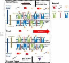
          Cell population heterogeneity can affect cellular response and is a major factor in drug resistance. However, there are few techniques available to represent and explore how heterogeneity is linked to population response. Recent high-throughput genomic, proteomic, and cellomic approaches offer opportunities for profiling heterogeneity on several scales. We have recently examined heterogeneity in vascular endothelial growth factor receptor (VEGFR) membrane localization in endothelial cells. We and others processed the heterogeneous data through ensemble averaging and integrated the data into computational models of anti-angiogenic drug effects in breast cancer. Here we show that additional modeling insight can be gained when cellular heterogeneity is considered. We present comprehensive statistical and computational methods for analyzing cellomic data sets and integrating them into deterministic models. We present a novel method for optimizing the fit of statistical distributions to heterogeneous data sets to preserve important data and exclude outliers. We compare methods of representing heterogeneous data and show methodology can affect model predictions up to 3.9-fold. We find that VEGF levels, a target for tuning angiogenesis, are more sensitive to VEGFR1 cell surface levels than VEGFR2; updating VEGFR1 levels in the tumor model gave a 64% change in free VEGF levels in the blood compartment, whereas updating VEGFR2 levels gave a 17% change. Furthermore, we find that subpopulations of tumor cells and tumor endothelial cells (tEC) expressing high levels of VEGFR (>35,000 VEGFR/cell) negate anti-VEGF treatments. We show that lowering the VEGFR membrane insertion rate for these subpopulations recovers the anti-angiogenic effect of anti-VEGF treatment, revealing new treatment targets for specific tumor cell subpopulations. This novel method of characterizing heterogeneous distributions shows for the first time how different representations of the same data set lead to different predictions of drug efficacy.

          Related collections

          Most cited references49

          • Record: found
          • Abstract: found
          • Article: not found

          Role of the vascular endothelial growth factor pathway in tumor growth and angiogenesis.

          New blood vessel formation (angiogenesis) is a fundamental event in the process of tumor growth and metastatic dissemination. Hence, the molecular basis of tumor angiogenesis has been of keen interest in the field of cancer research. The vascular endothelial growth factor (VEGF) pathway is well established as one of the key regulators of this process. The VEGF/VEGF-receptor axis is composed of multiple ligands and receptors with overlapping and distinct ligand-receptor binding specificities, cell-type expression, and function. Activation of the VEGF-receptor pathway triggers a network of signaling processes that promote endothelial cell growth, migration, and survival from pre-existing vasculature. In addition, VEGF mediates vessel permeability, and has been associated with malignant effusions. More recently, an important role for VEGF has emerged in mobilization of endothelial progenitor cells from the bone marrow to distant sites of neovascularization. The well-established role of VEGF in promoting tumor angiogenesis and the pathogenesis of human cancers has led to the rational design and development of agents that selectively target this pathway. Studies with various anti-VEGF/VEGF-receptor therapies have shown that these agents can potently inhibit angiogenesis and tumor growth in preclinical models. Recently, an anti-VEGF antibody (bevacizumab), when used in combination with chemotherapy, was shown to significantly improve survival and response rates in patients with metastatic colorectal cancer and thus, validate VEGF pathway inhibitors as an important new treatment modality in cancer therapy.
            Bookmark
            • Record: found
            • Abstract: found
            • Article: not found

            Vascular endothelial growth factor (VEGF) stimulates neurogenesis in vitro and in vivo.

            Vascular endothelial growth factor (VEGF) is an angiogenic protein with neurotrophic and neuroprotective effects. Because VEGF promotes the proliferation of vascular endothelial cells, we examined the possibility that it also stimulates the proliferation of neuronal precursors in murine cerebral cortical cultures and in adult rat brain in vivo. VEGF (>10 ng/ml) stimulated 5-bromo-2'-deoxyuridine (BrdUrd) incorporation into cells that expressed immature neuronal marker proteins and increased cell number in cultures by 20-30%. Cultured cells labeled by BrdUrd expressed VEGFR2/Flk-1, but not VEGFR1/Flt-1 receptors, and the effect of VEGF was blocked by the VEGFR2/Flk-1 receptor tyrosine kinase inhibitor SU1498. Intracerebroventricular administration of VEGF into rat brain increased BrdUrd labeling of cells in the subventricular zone (SVZ) and the subgranular zone (SGZ) of the hippocampal dentate gyrus (DG), where VEGFR2/Flk-1 was colocalized with the immature neuronal marker, doublecortin (Dcx). The increase in BrdUrd labeling after the administration of VEGF was caused by an increase in cell proliferation, rather than a decrease in cell death, because VEGF did not reduce caspase-3 cleavage in SVZ or SGZ. Cells labeled with BrdUrd after VEGF treatment in vivo include immature and mature neurons, astroglia, and endothelial cells. These findings implicate the angiogenesis factor VEGF in neurogenesis as well.
              Bookmark
              • Record: found
              • Abstract: found
              • Article: not found

              Heterogeneity of the tumor vasculature.

              The blood vessels supplying tumors are strikingly heterogeneous and differ from their normal counterparts with respect to organization, structure, and function. Six distinctly different tumor vessel types have been identified, and much has been learned about the steps and mechanisms by which they form. Four of the six vessel types (mother vessels, capillaries, glomeruloid microvascular proliferations, and vascular malformations) develop from preexisting normal venules and capillaries by angiogenesis. The two remaining vessel types (feeder arteries and draining veins) develop from arterio-venogenesis, a parallel, poorly understood process that involves the remodeling of preexisting arteries and veins. All six of these tumor vessel types can be induced to form sequentially in normal mouse tissues by an adenoviral vector expressing vascular endothelial growth factor (VEGF)-A164. Current antiangiogenic cancer therapies directed at VEGF-A or its receptors have been of only limited benefit to cancer patients, perhaps because they target only the endothelial cells of the tumor blood vessel subset that requires exogenous VEGF-A for maintenance. A goal of future work is to identify therapeutic targets on tumor blood vessel endothelial cells that have lost this requirement. Thieme Medical Publishers.
                Bookmark

                Author and article information

                Contributors
                Role: Editor
                Journal
                PLoS One
                PLoS ONE
                plos
                plosone
                PLoS ONE
                Public Library of Science (San Francisco, USA )
                1932-6203
                2014
                14 May 2014
                : 9
                : 5
                : e97271
                Affiliations
                [1]Department of Bioengineering, University of Illinois Urbana Champaign, Urbana, Illinois, United States of America
                Southern Illinois University School of Medicine, United States of America
                Author notes

                Competing Interests: The authors have declared that no competing interests exist.

                Conceived and designed the experiments: JW PII. Performed the experiments: JW. Analyzed the data: JW PII. Contributed reagents/materials/analysis tools: JW PII. Wrote the paper: JW.

                Article
                PONE-D-14-01050
                10.1371/journal.pone.0097271
                4020774
                24827582
                a6c0b74c-c498-4093-9ae6-55de1241b9fe
                Copyright @ 2014

                This is an open-access article distributed under the terms of the Creative Commons Attribution License, which permits unrestricted use, distribution, and reproduction in any medium, provided the original author and source are credited.

                History
                : 8 January 2014
                : 16 April 2014
                Page count
                Pages: 18
                Funding
                This work was supported by:NIH National Cancer Institute Research Supplements to Promote Diversity; American Cancer Society, Illinois Division Basic Research Grant. The funders had no role in study design, data collection and analysis, decision to publish, or preparation of the manuscript.
                Categories
                Research Article
                Biology and Life Sciences
                Anatomy
                Biological Tissue
                Epithelium
                Epithelial Cells
                Endothelial Cells
                Cell Biology
                Cell Motility
                Cell Migration
                Signal Transduction
                Cell Signaling
                Transmembrane Signaling
                Cellular Types
                Molecular Cell Biology
                Computational Biology
                Developmental Biology
                Molecular Development
                Morphogenesis
                Systems Biology
                Computer and Information Sciences
                Network Analysis
                Signaling Networks

                Uncategorized
                Uncategorized

                Comments

                Comment on this article