1
views
0
recommends
+1 Recommend
0 collections
    0
    shares
      • Record: found
      • Abstract: found
      • Article: found
      Is Open Access

      Skeletal Muscle Mass Reduction Velocity as a Simple Prognostic Indicator for Patients with Metastatic Urothelial Carcinoma Receiving Second-Line Chemotherapy

      research-article

      Read this article at

      Bookmark
          There is no author summary for this article yet. Authors can add summaries to their articles on ScienceOpen to make them more accessible to a non-specialist audience.

          Abstract

          Background:

          Patients with metastatic urothelial carcinoma (mUC) have an uncertain prognosis. The aim of the current study was to evaluate the prognostic potential of a skeletal muscle mass reduction index measured by computed tomography (CT) for mUC patients undergoing second-line gemcitabine and docetaxel (GD) chemotherapy.

          Methods:

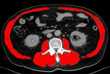
          We retrospectively reviewed 44 patients with mUC who received second-line GD chemotherapy between 2006 and 2015 in our hospital. Skeletal muscle area (SMA) at the third lumbar vertebra was measured using CT images obtained from medical records, and a skeletal muscle index (SMI) was calculated for each patient as: SMI = SMA / height2. Changes in SMI across timepoints (SMI inclination) were calculated as: SMI inclination = [( SMI/SMI)/duration of the interval between imaging visits]. Patients were then divided into two groups: a “steep” group (SMI inclination < -0.01) and a “gentle” group (SMI inclination ≥ -0.01). Kaplan-Meier curves and multivariate Cox proportional hazards regression models were used to evaluate the relationship between SMI inclination and overall survival (OS).

          Results:

          There were no differences in patient characteristics between the two groups with respect to median age, gender, Eastern Cooperative Oncology Group Performance Status (ECOG-PS), disease control rate or first-line treatment regimen. OS from the start of second-line GD therapy group was significantly shorter in the “steep” group relative to the “gentle” group. The multivariate analysis revealed that “steep” SMI inclination and presence of anemia were strong predictors of poor prognosis.

          Conclusion:

          Higher values of SMI inclination, indicating a faster rate of skeletal muscle mass reduction, may serve as a useful predictive marker for OS in mUC patients undergoing second-line GD chemotherapy.

          Related collections

          Most cited references17

          • Record: found
          • Abstract: found
          • Article: not found

          New response evaluation criteria in solid tumours: revised RECIST guideline (version 1.1).

          Assessment of the change in tumour burden is an important feature of the clinical evaluation of cancer therapeutics: both tumour shrinkage (objective response) and disease progression are useful endpoints in clinical trials. Since RECIST was published in 2000, many investigators, cooperative groups, industry and government authorities have adopted these criteria in the assessment of treatment outcomes. However, a number of questions and issues have arisen which have led to the development of a revised RECIST guideline (version 1.1). Evidence for changes, summarised in separate papers in this special issue, has come from assessment of a large data warehouse (>6500 patients), simulation studies and literature reviews. HIGHLIGHTS OF REVISED RECIST 1.1: Major changes include: Number of lesions to be assessed: based on evidence from numerous trial databases merged into a data warehouse for analysis purposes, the number of lesions required to assess tumour burden for response determination has been reduced from a maximum of 10 to a maximum of five total (and from five to two per organ, maximum). Assessment of pathological lymph nodes is now incorporated: nodes with a short axis of 15 mm are considered measurable and assessable as target lesions. The short axis measurement should be included in the sum of lesions in calculation of tumour response. Nodes that shrink to <10mm short axis are considered normal. Confirmation of response is required for trials with response primary endpoint but is no longer required in randomised studies since the control arm serves as appropriate means of interpretation of data. Disease progression is clarified in several aspects: in addition to the previous definition of progression in target disease of 20% increase in sum, a 5mm absolute increase is now required as well to guard against over calling PD when the total sum is very small. Furthermore, there is guidance offered on what constitutes 'unequivocal progression' of non-measurable/non-target disease, a source of confusion in the original RECIST guideline. Finally, a section on detection of new lesions, including the interpretation of FDG-PET scan assessment is included. Imaging guidance: the revised RECIST includes a new imaging appendix with updated recommendations on the optimal anatomical assessment of lesions. A key question considered by the RECIST Working Group in developing RECIST 1.1 was whether it was appropriate to move from anatomic unidimensional assessment of tumour burden to either volumetric anatomical assessment or to functional assessment with PET or MRI. It was concluded that, at present, there is not sufficient standardisation or evidence to abandon anatomical assessment of tumour burden. The only exception to this is in the use of FDG-PET imaging as an adjunct to determination of progression. As is detailed in the final paper in this special issue, the use of these promising newer approaches requires appropriate clinical validation studies.
            Bookmark
            • Record: found
            • Abstract: found
            • Article: not found

            Sarcopenia: European consensus on definition and diagnosis

            The European Working Group on Sarcopenia in Older People (EWGSOP) developed a practical clinical definition and consensus diagnostic criteria for age-related sarcopenia. EWGSOP included representatives from four participant organisations, i.e. the European Geriatric Medicine Society, the European Society for Clinical Nutrition and Metabolism, the International Association of Gerontology and Geriatrics—European Region and the International Association of Nutrition and Aging. These organisations endorsed the findings in the final document. The group met and addressed the following questions, using the medical literature to build evidence-based answers: (i) What is sarcopenia? (ii) What parameters define sarcopenia? (iii) What variables reflect these parameters, and what measurement tools and cut-off points can be used? (iv) How does sarcopenia relate to cachexia, frailty and sarcopenic obesity? For the diagnosis of sarcopenia, EWGSOP recommends using the presence of both low muscle mass + low muscle function (strength or performance). EWGSOP variously applies these characteristics to further define conceptual stages as ‘presarcopenia’, ‘sarcopenia’ and ‘severe sarcopenia’. EWGSOP reviewed a wide range of tools that can be used to measure the specific variables of muscle mass, muscle strength and physical performance. Our paper summarises currently available data defining sarcopenia cut-off points by age and gender; suggests an algorithm for sarcopenia case finding in older individuals based on measurements of gait speed, grip strength and muscle mass; and presents a list of suggested primary and secondary outcome domains for research. Once an operational definition of sarcopenia is adopted and included in the mainstream of comprehensive geriatric assessment, the next steps are to define the natural course of sarcopenia and to develop and define effective treatment.
              Bookmark
              • Record: found
              • Abstract: found
              • Article: not found

              Definition and classification of cancer cachexia: an international consensus.

              To develop a framework for the definition and classification of cancer cachexia a panel of experts participated in a formal consensus process, including focus groups and two Delphi rounds. Cancer cachexia was defined as a multifactorial syndrome defined by an ongoing loss of skeletal muscle mass (with or without loss of fat mass) that cannot be fully reversed by conventional nutritional support and leads to progressive functional impairment. Its pathophysiology is characterised by a negative protein and energy balance driven by a variable combination of reduced food intake and abnormal metabolism. The agreed diagnostic criterion for cachexia was weight loss greater than 5%, or weight loss greater than 2% in individuals already showing depletion according to current bodyweight and height (body-mass index [BMI] <20 kg/m(2)) or skeletal muscle mass (sarcopenia). An agreement was made that the cachexia syndrome can develop progressively through various stages--precachexia to cachexia to refractory cachexia. Severity can be classified according to degree of depletion of energy stores and body protein (BMI) in combination with degree of ongoing weight loss. Assessment for classification and clinical management should include the following domains: anorexia or reduced food intake, catabolic drive, muscle mass and strength, functional and psychosocial impairment. Consensus exists on a framework for the definition and classification of cancer cachexia. After validation, this should aid clinical trial design, development of practice guidelines, and, eventually, routine clinical management. Copyright © 2011 Elsevier Ltd. All rights reserved.
                Bookmark

                Author and article information

                Journal
                Asian Pac J Cancer Prev
                Asian Pac. J. Cancer Prev
                APJCP
                Asian Pacific Journal of Cancer Prevention : APJCP
                West Asia Organization for Cancer Prevention (Iran )
                1513-7368
                2476-762X
                2019
                : 20
                : 10
                : 2995-3000
                Affiliations
                [1 ] Department of Nephro‐Urology, Nagoya City University, Graduate School of Medical Sciences,
                [3 ] Department of Pharmacy, Nagoya City University Hospital, Nagoya,
                [2 ] Department of Urology, Anjo Kosei Hospital, Anjo City, Japan.
                Author notes
                [* ]For Correspondence: naiki@med.nagoya-cu.ac.jp
                Article
                10.31557/APJCP.2019.20.10.2995
                6982681
                31653146
                a7c096bb-a214-459f-ba25-75199b0f6f07

                This is an Open Access article distributed under the terms of the Creative Commons Attribution License, ( http://creativecommons.org/licenses/by/3.0/) which permits unrestricted use, distribution, and reproduction in any medium, provided the original work is properly cited.

                History
                : 8 May 2019
                : 22 September 2019
                Categories
                Research Article

                metastatic urothelial carcinoma,second-line chemotherapy,prognostic factor,sarcopenia

                Comments

                Comment on this article

                scite_

                Similar content151

                Cited by3

                Most referenced authors561