21
views
0
recommends
+1 Recommend
0 collections
    0
    shares
      • Record: found
      • Abstract: found
      • Article: found
      Is Open Access

      KCa3.1 mediates dysfunction of tubular autophagy in diabetic kidneys via PI3k/Akt/mTOR signaling pathways

      research-article

      Read this article at

      Bookmark
          There is no author summary for this article yet. Authors can add summaries to their articles on ScienceOpen to make them more accessible to a non-specialist audience.

          Abstract

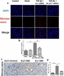
          Autophagy is emerging as an important pathway in many diseases including diabetic nephropathy. It is acknowledged that oxidative stress plays a critical role in autophagy dysfunction and diabetic nephropathy, and KCa3.1 blockade ameliorates diabetic renal fibrosis through inhibiting TGF-β1 signaling pathway. To identify the role of KCa3.1 in dysfunctional tubular autophagy in diabetic nephropathy, human proximal tubular cells (HK2) transfected with scrambled or KCa3.1 siRNAs were exposed to TGF-β1 for 48 h, then autophagosome formation, the autophagy marker LC3, signaling molecules PI3K, Akt and mTOR, and oxidative stress marker nitrotyrosine were examined respectively. In vivo, LC3, nitrotyrosine and phosphorylated mTOR were examined in kidneys of diabetic KCa3.1+/+ and KCa3.1−/− mice. The results demonstrated that TGF-β1 increased the formation of autophagic vacuoles, LC3 expression, and phosphorylation of PI3K, Akt and mTOR in scrambled siRNA transfected HK2 cells compared to control cells, which was reversed in KCa3.1 siRNA transfected HK2 cells. In vivo, expression of LC3 and nitrotyrosine, and phosphorylation of mTOR were significantly increased in kidneys of diabetic KCa3.1+/+ mice compared to non-diabetic mice, which were attenuated in kidneys of diabetic KCa3.1−/− mice. These results suggest that KCa3.1 activation contributes to dysfunctional tubular autophagy in diabetic nephropathy through PI3K/Akt/mTOR signaling pathways.

          Related collections

          Most cited references30

          • Record: found
          • Abstract: found
          • Article: not found

          The thioredoxin system as a therapeutic target in human health and disease.

          The thioredoxin (Trx) system comprises Trx, truncated Trx (Trx-80), Trx reductase, and NADPH, besides a natural Trx inhibitor, the thioredoxin-interacting protein (TXNIP). This system is essential for maintaining the balance of the cellular redox status, and it is involved in the regulation of redox signaling. It is also pivotal for growth promotion, neuroprotection, inflammatory modulation, antiapoptosis, immune function, and atherosclerosis. As an ubiquitous and multifunctional protein, Trx is expressed in all forms of life, executing its function through its antioxidative, protein-reducing, and signal-transducing activities. In this review, the biological properties of the Trx system are highlighted, and its implications in several human diseases are discussed, including cardiovascular diseases, heart failure, stroke, inflammation, metabolic syndrome, neurodegenerative diseases, arthritis, and cancer. The last chapter addresses the emerging therapeutic approaches targeting the Trx system in human diseases.
            Bookmark
            • Record: found
            • Abstract: found
            • Article: not found

            Up-regulation of the IKCa1 potassium channel during T-cell activation. Molecular mechanism and functional consequences.

            We used whole cell recording to evaluate functional expression of the intermediate conductance Ca(2+)-activated K(+) channel, IKCa1, in response to various mitogenic stimuli. One to two days following engagement of T-cell receptors to trigger both PKC- and Ca(2+)-dependent events, IKCa1 expression increased from an average of 8 to 300-800 channels/cell. Selective stimulation of the PKC pathway resulted in equivalent up-regulation, whereas a calcium ionophore was relatively ineffective. Enhancement in IKCa1 mRNA levels paralleled the increased channel number. The genomic organization of IKCa1, SKCa2, and SKCa3 were defined, and IK(Ca) and SK(Ca) genes were found to have a remarkably similar intron-exon structure. Mitogens enhanced IKCa1 promoter activity proportional to the increase in IKCa1 mRNA, suggesting that transcriptional mechanisms underlie channel up-regulation. Mutation of motifs for AP1 and Ikaros-2 in the promoter abolished this induction. Selective Kv1.3 inhibitors ShK-Dap(22), margatoxin, and correolide suppressed mitogenesis of resting T-cells but not preactivated T-cells with up-regulated IKCa1 channel expression. Selectively blocking IKCa1 channels with clotrimazole or TRAM-34 suppressed mitogenesis of preactivated lymphocytes, whereas resting T-cells were less sensitive. Thus, Kv1.3 channels are essential for activation of quiescent cells, but signaling through the PKC pathway enhances expression of IKCa1 channels that are required for continued proliferation.
              Bookmark
              • Record: found
              • Abstract: found
              • Article: found
              Is Open Access

              TXNIP Links Innate Host Defense Mechanisms to Oxidative Stress and Inflammation in Retinal Muller Glia under Chronic Hyperglycemia: Implications for Diabetic Retinopathy

              Thioredoxin Interacting Protein (TXNIP) mediates retinal inflammation, gliosis, and apoptosis in experimental diabetes. Here, we investigate the temporal response of Muller glia to high glucose (HG) and TXNIP expression using a rat Muller cell line (rMC1) in culture. We examined if HG-induced TXNIP expression evokes host defense mechanisms in rMC1 in response to metabolic abnormalities. HG causes sustained up-regulation of TXNIP (2 h to 5 days), ROS generation, ATP depletion, ER stress, and inflammation. Various cellular defense mechanisms are activated by HG: (i) NLRP3 inflammasome, (ii) ER stress response (sXBP1), (iii) hypoxic-like HIF-1 α induction, (iv) autophagy/mitophagy, and (v) apoptosis. We also found in vivo that streptozocin-induced diabetic rats have higher retinal TXNIP and innate immune response gene expression than normal rats. Knock down of TXNIP by intravitreal siRNA reduces inflammation (IL-1 β ) and gliosis (GFAP) in the diabetic retina. TXNIP ablation in vitro prevents ROS generation, restores ATP level and autophagic LC3B induction in rMC1. Thus, our results show that HG sustains TXNIP up-regulation in Muller glia and evokes a program of cellular defense/survival mechanisms that ultimately lead to oxidative stress, ER stress/inflammation, autophagy and apoptosis. TXNIP is a potential target to ameliorate blinding ocular complications of diabetic retinopathy.
                Bookmark

                Author and article information

                Journal
                Sci Rep
                Sci Rep
                Scientific Reports
                Nature Publishing Group
                2045-2322
                31 March 2016
                2016
                : 6
                : 23884
                Affiliations
                [1 ]Kolling Institute of Medical Research, Sydney Medical School, University of Sydney, Royal North Shore Hospital, St Leonards , Sydney, NSW 2065, Australia
                [2 ]School of Medical Sciences (Discipline of Anatomy and Histology) – The Bosch Institute, University of Sydney , NSW 2006, Australia
                [3 ]Australian Centre for Microscopy & Microanalysis, Madsen Building, University of Sydney , NSW 2006, Australia
                Author notes
                Article
                srep23884
                10.1038/srep23884
                4814925
                27029904
                a846d831-00f1-4cea-8789-8f2963061b56
                Copyright © 2016, Macmillan Publishers Limited

                This work is licensed under a Creative Commons Attribution 4.0 International License. The images or other third party material in this article are included in the article’s Creative Commons license, unless indicated otherwise in the credit line; if the material is not included under the Creative Commons license, users will need to obtain permission from the license holder to reproduce the material. To view a copy of this license, visit http://creativecommons.org/licenses/by/4.0/

                History
                : 20 July 2015
                : 16 February 2016
                Categories
                Article

                Uncategorized
                Uncategorized

                Comments

                Comment on this article