10
views
0
recommends
+1 Recommend
0 collections
    0
    shares
      • Record: found
      • Abstract: found
      • Article: found
      Is Open Access

      Artificial Intelligence for the Future Radiology Diagnostic Service

      review-article

      Read this article at

      Bookmark
          There is no author summary for this article yet. Authors can add summaries to their articles on ScienceOpen to make them more accessible to a non-specialist audience.

          Abstract

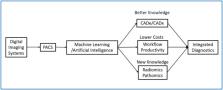
          Radiology historically has been a leader of digital transformation in healthcare. The introduction of digital imaging systems, picture archiving and communication systems (PACS), and teleradiology transformed radiology services over the past 30 years. Radiology is again at the crossroad for the next generation of transformation, possibly evolving as a one-stop integrated diagnostic service. Artificial intelligence and machine learning promise to offer radiology new powerful new digital tools to facilitate the next transformation. The radiology community has been developing computer-aided diagnosis (CAD) tools based on machine learning (ML) over the past 20 years. Among various AI techniques, deep-learning convolutional neural networks (CNN) and its variants have been widely used in medical image pattern recognition. Since the 1990s, many CAD tools and products have been developed. However, clinical adoption has been slow due to a lack of substantial clinical advantages, difficulties integrating into existing workflow, and uncertain business models. This paper proposes three pathways for AI's role in radiology beyond current CNN based capabilities 1) improve the performance of CAD, 2) improve the productivity of radiology service by AI-assisted workflow, and 3) develop radiomics that integrate the data from radiology, pathology, and genomics to facilitate the emergence of a new integrated diagnostic service.

          Related collections

          Most cited references59

          • Record: found
          • Abstract: found
          • Article: not found

          The Image Biomarker Standardization Initiative: Standardized Quantitative Radiomics for High-Throughput Image-based Phenotyping

          Background Radiomic features may quantify characteristics present in medical imaging. However, the lack of standardized definitions and validated reference values have hampered clinical use. Purpose To standardize a set of 174 radiomic features. Materials and Methods Radiomic features were assessed in three phases. In phase I, 487 features were derived from the basic set of 174 features. Twenty-five research teams with unique radiomics software implementations computed feature values directly from a digital phantom, without any additional image processing. In phase II, 15 teams computed values for 1347 derived features using a CT image of a patient with lung cancer and predefined image processing configurations. In both phases, consensus among the teams on the validity of tentative reference values was measured through the frequency of the modal value and classified as follows: less than three matches, weak; three to five matches, moderate; six to nine matches, strong; 10 or more matches, very strong. In the final phase (phase III), a public data set of multimodality images (CT, fluorine 18 fluorodeoxyglucose PET, and T1-weighted MRI) from 51 patients with soft-tissue sarcoma was used to prospectively assess reproducibility of standardized features. Results Consensus on reference values was initially weak for 232 of 302 features (76.8%) at phase I and 703 of 1075 features (65.4%) at phase II. At the final iteration, weak consensus remained for only two of 487 features (0.4%) at phase I and 19 of 1347 features (1.4%) at phase II. Strong or better consensus was achieved for 463 of 487 features (95.1%) at phase I and 1220 of 1347 features (90.6%) at phase II. Overall, 169 of 174 features were standardized in the first two phases. In the final validation phase (phase III), most of the 169 standardized features could be excellently reproduced (166 with CT; 164 with PET; and 164 with MRI). Conclusion A set of 169 radiomics features was standardized, which enabled verification and calibration of different radiomics software. © RSNA, 2020 Online supplemental material is available for this article. See also the editorial by Kuhl and Truhn in this issue.
            Bookmark
            • Record: found
            • Abstract: found
            • Article: not found

            Artificial intelligence in radiology

            Artificial intelligence (AI) algorithms, particularly deep learning, have demonstrated remarkable progress in image-recognition tasks. Methods ranging from convolutional neural networks to variational autoencoders have found myriad applications in the medical image analysis field, propelling it forward at a rapid pace. Historically, in radiology practice, trained physicians visually assessed medical images for the detection, characterization and monitoring of diseases. AI methods excel at automatically recognizing complex patterns in imaging data and providing quantitative, rather than qualitative, assessments of radiographic characteristics. In this O pinion article, we establish a general understanding of AI methods, particularly those pertaining to image-based tasks. We explore how these methods could impact multiple facets of radiology, with a general focus on applications in oncology, and demonstrate ways in which these methods are advancing the field. Finally, we discuss the challenges facing clinical implementation and provide our perspective on how the domain could be advanced.
              Bookmark
              • Record: found
              • Abstract: found
              • Article: found
              Is Open Access

              Convolutional neural networks: an overview and application in radiology

              Abstract Convolutional neural network (CNN), a class of artificial neural networks that has become dominant in various computer vision tasks, is attracting interest across a variety of domains, including radiology. CNN is designed to automatically and adaptively learn spatial hierarchies of features through backpropagation by using multiple building blocks, such as convolution layers, pooling layers, and fully connected layers. This review article offers a perspective on the basic concepts of CNN and its application to various radiological tasks, and discusses its challenges and future directions in the field of radiology. Two challenges in applying CNN to radiological tasks, small dataset and overfitting, will also be covered in this article, as well as techniques to minimize them. Being familiar with the concepts and advantages, as well as limitations, of CNN is essential to leverage its potential in diagnostic radiology, with the goal of augmenting the performance of radiologists and improving patient care. Key Points • Convolutional neural network is a class of deep learning methods which has become dominant in various computer vision tasks and is attracting interest across a variety of domains, including radiology. • Convolutional neural network is composed of multiple building blocks, such as convolution layers, pooling layers, and fully connected layers, and is designed to automatically and adaptively learn spatial hierarchies of features through a backpropagation algorithm. • Familiarity with the concepts and advantages, as well as limitations, of convolutional neural network is essential to leverage its potential to improve radiologist performance and, eventually, patient care.
                Bookmark

                Author and article information

                Contributors
                Journal
                Front Mol Biosci
                Front Mol Biosci
                Front. Mol. Biosci.
                Frontiers in Molecular Biosciences
                Frontiers Media S.A.
                2296-889X
                28 January 2021
                2020
                : 7
                : 614258
                Affiliations
                Arlington Innovation Center:Health Research, Virginia Tech-Washington DC Area, Arlington, VA, United States
                Author notes

                Edited by: Xin Gao, King Abdullah University of Science and Technology, Saudi Arabia

                Reviewed by: Salva Mena-Mollá, University of Valencia, Spain

                Mohammad Shahid, Children’s National Hospital, United States

                *Correspondence: Seong K. Mun, munsk@ 123456vt.edu

                This article was submitted to Molecular Diagnostics and Therapeutics, a section of the journal Frontiers in Molecular Biosciences

                Article
                614258
                10.3389/fmolb.2020.614258
                7875875
                33585563
                a88af677-6a1a-4c65-b2a9-a0e61fb027d9
                Copyright © 2021 Mun, Wong, Lo, Li and Bayarsaikhan.

                This is an open-access article distributed under the terms of the Creative Commons Attribution License (CC BY). The use, distribution or reproduction in other forums is permitted, provided the original author(s) and the copyright owner(s) are credited and that the original publication in this journal is cited, in accordance with accepted academic practice. No use, distribution or reproduction is permitted which does not comply with these terms.

                History
                : 05 October 2020
                : 29 December 2020
                Categories
                Molecular Biosciences
                Review

                artificial intelligence,radiology,cnn,productivity,integrated diagnostics,workflow

                Comments

                Comment on this article