8
views
0
recommends
+1 Recommend
0 collections
    0
    shares
      • Record: found
      • Abstract: found
      • Article: found
      Is Open Access

      Biomarkers Associated With Aortic Valve Calcification: Should We Focus on Sex Specific Processes?

      research-article

      Read this article at

      Bookmark
          There is no author summary for this article yet. Authors can add summaries to their articles on ScienceOpen to make them more accessible to a non-specialist audience.

          Abstract

          Objective

          Circulating biomarkers are useful in detection and monitoring of cardiovascular diseases. However, their role in aortic valve disease is unclear. Mechanisms are rapidly elucidated and sex differences are suggested to be involved. Therefore, we sought to identify biomarkers involved in aortic valve calcification (AVC) stratified by sex.

          Methods

          Blood samples of 34 patients with AVC (without further overt cardiovascular disease, including absence of hemodynamic consequences of valvular calcification) were compared with 136 patients without AVC. AVC was determined using computed tomography calcium scoring. Circulating biomarkers were quantified using a novel antibody-based method (Olink Proseek Multiplex Cardiovascular Panel I) and 92 biomarkers were compared between patients with and without AVC.

          Results

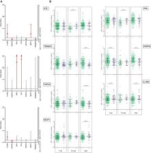
          In the overall population, Interleukin-1 Receptor Antagonist and pappalysin-1 were associated with increased and decreased odds of having AVC. These differences were driven by the male population [IL1RA: OR 2.79 (1.16–6.70), p = 0.022; PAPPA: OR 0.30 (0.11–0.84), p = 0.021]. Furthermore, TNF-related activation-induced cytokine (TRANCE) and fibroblast growth factor-23 were associated decreased odds of having AVC, and monocyte chemotactic protein-1 was associated with increased odds of having AVC [TRANCE: OR 0.32 (0.12–0.80), p = 0.015; FGF23: OR 0.41 (0.170–0.991), p = 0.048; MCP1: OR 2.64 (1.02–6.81), p = 0.045]. In contrast, galanin peptides and ST2 were associated with increased odds of having AVC in females [GAL: OR 12.38 (1.31–116.7), p = 0.028; ST2: OR13.64 (1.21–153.33), p = 0.034].

          Conclusion

          In this exploratory study, we identified biomarkers involved in inflammation, fibrosis and calcification which may be associated with having AVC. Biomarkers involved in fibrosis may show higher expression in females, whilst biomarkers involved in inflammation and calcification could associate with AVC in males.

          Related collections

          Most cited references27

          • Record: found
          • Abstract: found
          • Article: not found

          Characterization of the early lesion of 'degenerative' valvular aortic stenosis. Histological and immunohistochemical studies.

          Nonrheumatic stenosis of trileaflet aortic valves, often termed senile or calcific valvular aortic stenosis, is considered a "degenerative" process, but little is known about the cellular or molecular factors that mediate its development. To characterize the developing aortic valvular lesion, we performed histological and immunohistochemical studies on Formalin-fixed and methanol-Carnoy's-fixed paraffin-embedded aortic valve leaflets or on frozen sections obtained at autopsy from 27 adults (age, 46 to 82 years) with normal leaflets (n = 6), mild macroscopic leaflet thickening (n = 15), or clinical aortic stenosis (n = 6). Focal areas of thickening ("early lesions") were characterized by (1) subendothelial thickening on the aortic side of the leaflet, between the basement membrane (PAS-positive) and elastic lamina (Verhoeff-van Gieson), (2) the presence of large amounts of intracellular and extracellular neutral lipids (oil red O) and fine, stippled mineralization (von Kossa), and (3) disruption of the basement membrane overlying the lesion. Regions of the fibrosa adjacent to these lesions were characterized by thickening and by protein, lipid, and calcium accumulation. Control valves showed none of these abnormalities. Immunohistochemical studies were performed using monoclonal antibodies directed against macrophages (anti-CD68 or HAM-56), and contractile proteins of smooth muscle cells or myofibroblasts (anti-alpha-actin and HHF-35) or rabbit polyclonal antiserum against T lymphocytes (anti-CD3). In normal valves, scattered macrophages were present in the fibrosa and ventricularis, and occasional muscle actin-positive cells were detected in the proximal portion of the ventricularis near the leaflet base, but no T lymphocytes were found. In contrast, early lesions were characterized by the presence of an inflammatory infiltrate composed of non-foam cell and foam cell macrophages, occasional T cells, and rare alpha-actin-positive cells. In stenotic aortic valves, a similar but more advanced lesion was seen. The early lesion of "degenerative" aortic stenosis is an active inflammatory process with some similarities (lipid deposition, macrophage and T-cell infiltration, and basement membrane disruption) and some dissimilarities (presence of prominent mineralization and small numbers of smooth muscle cells) to atherosclerosis.
            Bookmark
            • Record: found
            • Abstract: found
            • Article: not found

            Calcification in Aortic Stenosis: The Skeleton Key.

            Aortic stenosis is a common, potentially fatal condition that is set to become an increasing public health burden. Once symptoms develop, there is an inexorable deterioration with a poor prognosis. Despite this, there are no medical therapies capable of modifying disease progression, and the only available treatment is aortic valve replacement, to which not all patients are suited. Conventional teaching suggests that aortic stenosis is a degenerative condition whereby "wear and tear" leads to calcium deposition within the valve. Although mechanical stress and injury are important factors, it is becoming increasingly appreciated that aortic stenosis is instead governed by a highly complex, regulated pathological process with similarities to skeletal bone formation. This review discusses the pathophysiology of aortic stenosis with an emphasis on the emerging importance of calcification, how this can be visualized and monitored using noninvasive imaging, and how our improved knowledge may ultimately translate into novel disease-modifying treatments.
              Bookmark
              • Record: found
              • Abstract: found
              • Article: not found

              Impact of aortic valve calcification, as measured by MDCT, on survival in patients with aortic stenosis: results of an international registry study.

              Aortic valve calcification (AVC) load measures lesion severity in aortic stenosis (AS) and is useful for diagnostic purposes. Whether AVC predicts survival after diagnosis, independent of clinical and Doppler echocardiographic AS characteristics, has not been studied.
                Bookmark

                Author and article information

                Contributors
                Journal
                Front Cell Dev Biol
                Front Cell Dev Biol
                Front. Cell Dev. Biol.
                Frontiers in Cell and Developmental Biology
                Frontiers Media S.A.
                2296-634X
                10 July 2020
                2020
                : 8
                : 604
                Affiliations
                [1] 1Department of Cardiology and CARIM, Maastricht University Medical Center+, School for Cardiovascular Diseases , Maastricht, Netherlands
                [2] 2Institute of Cardiovascular Sciences, University of Birmingham , Birmingham, United Kingdom
                [3] 3Department of Radiology and Nuclear Medicine and CARIM, Maastricht University Medical Center+, School for Cardiovascular Diseases , Maastricht, Netherlands
                [4] 4Department of Clinical Chemistry and CARIM, Maastricht University Medical Center+, School for Cardiovascular Diseases , Maastricht, Netherlands
                [5] 5Department of Biochemistry and CARIM, Maastricht University, School for Cardiovascular Diseases , Maastricht, Netherlands
                Author notes

                Edited by: Herve Kempf, UMR 7365 Ingénierie Moléculaire et Physiopathologie Articulaire (IMOPA), France

                Reviewed by: Gonzalo Artiach, Karolinska University Hospital, Sweden; Noah Lucas Weisleder, The Ohio State University, United States

                *Correspondence: Frederique E. C. M. Peeters, frederique.peeters@ 123456mumc.nl

                Present address: Bas L. J. H. Kietselaer, Zuyderland Medisch Centrum, Heerlen, Netherlands

                This article was submitted to Molecular Medicine, a section of the journal Frontiers in Cell and Developmental Biology

                Article
                10.3389/fcell.2020.00604
                7366171
                32754594
                a8e31d29-f498-4cfb-a7dd-cc3784471a59
                Copyright © 2020 Peeters, Dudink, Weijs, Fabritz, Chua, Kietselaer, Wildberger, Meex, Kirchhof, Crijns and Schurgers.

                This is an open-access article distributed under the terms of the Creative Commons Attribution License (CC BY). The use, distribution or reproduction in other forums is permitted, provided the original author(s) and the copyright owner(s) are credited and that the original publication in this journal is cited, in accordance with accepted academic practice. No use, distribution or reproduction is permitted which does not comply with these terms.

                History
                : 28 February 2020
                : 19 June 2020
                Page count
                Figures: 1, Tables: 1, Equations: 0, References: 40, Pages: 9, Words: 0
                Categories
                Cell and Developmental Biology
                Original Research

                aortic valve calcification,biomarkers,sex-specific,fibrosis,inflammation

                Comments

                Comment on this article