41
views
0
recommends
+1 Recommend
0 collections
    1
    shares
      • Record: found
      • Abstract: found
      • Article: found
      Is Open Access

      Beneficial effects of a novel shark-skin collagen dressing for the promotion of seawater immersion wound healing

      research-article

      Read this article at

      Bookmark
          There is no author summary for this article yet. Authors can add summaries to their articles on ScienceOpen to make them more accessible to a non-specialist audience.

          Abstract

          Background

          Wounded personnel who work at sea often encounter a plethora of difficulties. The most important of these difficulties is seawater immersion. Common medical dressings have little effect when the affected area is immersed in seawater, and only rarely dressings have been reported for the treatment of seawater-immersed wounds. The objective of this study is to develop a new dressing which should be suitable to prevent the wound from seawater immersion and to promote the wound healing.

          Methods

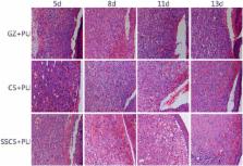
          Shark skin collagen (SSC) was purified via ethanol de-sugaring and de-pigmentation and adjusted for pH. A shark skin collagen sponge (SSCS) was prepared by freeze-drying. SSCS was attached to an anti-seawater immersion polyurethane (PU) film (SSCS + PU) to compose a new dressing. The biochemical properties of SSC and physicochemical properties of SSCS were assessed by standard methods. The effects of SSCS and SSCS + PU on the healing of seawater-immersed wounds were studied using a seawater immersion rat model. For the detection of SSCS effects on seawater-immersed wounds, 12 SD rats, with four wounds created in each rat, were divided into four groups: the 3rd day group, 5th day group, 7th day group and 12th day group. In each group, six wounds were treated with SSCS, three wounds treated with chitosan served as the positive control, and three wounds treated with gauze served as the negative control. For the detection of the SSCS + PU effects on seawater-immersed wounds, 36 SD rats were divided into three groups: the gauze (GZ) + PU group, chitosan (CS) + PU group and SSCS + PU group, with 12 rats in each group, and two wounds in each rat. The wound sizes were measured to calculate the healing rate, and histomorphology and the immunohistochemistry of the CD31 and TGF-β expression levels in the wounded tissues were measured by standard methods.

          Results

          The results of Ultraviolet-visible (UV-vis) spectrum, Fourier-transform infrared (FTIR) spectrum, circular dichroism (CD) spectra, sodium dodecyl sulfate polyacrylamide gel electrophoresis (SDS-PAGE), and amino acid composition analyses of SSC demonstrated that SSC is type I collagen. SSCS had a homogeneous porous structure of approximately 200 μm, porosity rate of 83.57% ± 2.64%, water vapor transmission ratio (WVTR) of 4500 g/m 2, tensile strength of 1.79 ± 0.41 N/mm, and elongation at break of 4.52% ± 0.01%. SSCS had significant beneficial effects on seawater-immersed wound healing. On the 3rd day, the healing rates in the GZ negative control, CS positive control and SSCS rats were 13.94% ± 5.50%, 29.40% ± 1.10% and 47.24% ± 8.40%, respectively. SSCS also enhanced TGF-β and CD31 expression in the initial stage of the healing period. The SSCS + PU dressing effectively protected wounds from seawater immersion for at least 4 h, and accelerated re-epithelialization, vascularization and granulation formation of seawater-immersed wounds in the earlier stages of wound healing, and as well as significantly promoted wound healing. The SSCS + PU dressing also enhanced expression of TGF-β and CD31. The effects of SSCS and SSCS + PU were superior to those of both the chitosan and gauze dressings.

          Conclusions

          SSCS has significant positive effects on the promotion of seawater-immersed wound healing, and a SSCS + PU dressing effectively prevents seawater immersion, and significantly promotes seawater-immersed wound healing.

          Related collections

          Most cited references29

          • Record: found
          • Abstract: found
          • Article: not found

          Signaling pathway of MAPK/ERK in cell proliferation, differentiation, migration, senescence and apoptosis.

          The generic mitogen-activated protein kinases (MAPK) signaling pathway is shared by four distinct cascades, including the extracellular signal-related kinases (ERK1/2), Jun amino-terminal kinases (JNK1/2/3), p38-MAPK and ERK5. Mitogen-activated protein kinases/extracellular signal-regulated kinase (MAPK/ERK) pathway is reported to be associated with the cell proliferation, differentiation, migration, senescence and apoptosis. The literatures were searched extensively and this review was performed to review the role of MAPK/ERK signaling pathway in cell proliferation, differentiation, migration, senescence and apoptosis.
            Bookmark
            • Record: found
            • Abstract: found
            • Article: not found

            Critical Role of Transforming Growth Factor Beta in Different Phases of Wound Healing.

            This review highlights the critical role of transforming growth factor beta (TGF-β)1-3 within different phases of wound healing, in particular, late-stage wound healing. It is also very important to identify the TGF-β1-controlling factors involved in slowing down the healing process upon wound epithelialization. TGF-β1, as a growth factor, is a known proponent of dermal fibrosis. Several strategies to modulate or regulate TGF's actions have been thoroughly investigated in an effort to create successful therapies. This study reviews current discourse regarding the many roles of TGF-β1 in wound healing by modulating infiltrated immune cells and the extracellular matrix. It is well established that TGF-β1 functions as a wound-healing promoting factor, and thereby if in excess it may lead to overhealing outcomes, such as hypertrophic scarring and keloid. Thus, the regulation of TGF-β1 in the later stages of the healing process remains as critical issue of which to better understand. One hypothesis is that cell communication is the key to regulate later stages of wound healing. To elucidate the role of keratinocyte/fibroblast cross talk in controlling the later stages of wound healing we need to: (1) identify those keratinocyte-released factors which would function as wound-healing stop signals, (2) evaluate the functionality of these factors in controlling the outcome of the healing process, and (3) formulate topical vehicles for these antifibrogenic factors to improve or even prevent the development of hypertrophic scarring and keloids as a result of deep trauma, burn injuries, and any type of surgical incision.
              Bookmark
              • Record: found
              • Abstract: found
              • Article: not found

              Signaling by members of the TGF-beta family in vascular morphogenesis and disease.

              Members of the transforming growth factor-beta (TGF-beta) family play pivotal roles in development and disease. These cytokines elicit their pleiotropic effects on cells, including endothelial and mural cells, through specific type I and type II serine/threonine kinase receptors and intracellular Smad transcription factors. This review highlights recent progress in our understanding of TGF-beta signaling in vascular development and angiogenesis and of how perturbed TGF-beta signaling might contribute to vascular pathologies, tumor angiogenesis and tumor progression. Recent research has provided exciting insights into the role of the TGF-beta type I receptor (ALK1) in tumor angiogenesis and the curative effects of thalidomide on vascular malformations in hereditary hemorrhagic telangiectasia (HHT). These advances provide opportunities for the development of new therapies for diseases with vascular abnormalities. Copyright 2010 Elsevier Ltd. All rights reserved.
                Bookmark

                Author and article information

                Contributors
                xianrong_sh@163.com
                cxl219912@163.com
                xhx7760@163.com
                yinghe_hys@163.com
                vip2050@sina.com
                qun246013@163.com
                379084935@qq.com
                373190610@qq.com
                houdy2@163.com
                jiangdingwen@sina.com
                qingrongw@126.com
                Journal
                Mil Med Res
                Mil Med Res
                Military Medical Research
                BioMed Central (London )
                2095-7467
                2054-9369
                27 October 2017
                27 October 2017
                2017
                : 4
                : 33
                Affiliations
                [1 ]ISNI 0000 0004 1755 2063, GRID grid.415934.e, The PLA Key Laboratory of Biological Effect and Medical Protection on Naval Vessel Special Environment, Naval Medical Research Institute, ; Shanghai, 200433 China
                [2 ]ISNI 0000 0000 9833 2433, GRID grid.412514.7, College of Food Science and Technology, Shanghai Ocean University, ; Shanghai, 201306 China
                [3 ]ISNI 0000 0000 8744 8924, GRID grid.268505.c, Research Center of TCM Processing Technology, , Zhejiang Chinese Medical University, ; Hang Zhou, 311401 China
                Author information
                http://orcid.org/0000-0001-6207-5305
                Article
                143
                10.1186/s40779-017-0143-4
                5658943
                29502521
                a9ee23c3-f2d4-4335-a91a-acdec102a380
                © The Author(s). 2017

                Open AccessThis article is distributed under the terms of the Creative Commons Attribution 4.0 International License ( http://creativecommons.org/licenses/by/4.0/), which permits unrestricted use, distribution, and reproduction in any medium, provided you give appropriate credit to the original author(s) and the source, provide a link to the Creative Commons license, and indicate if changes were made. The Creative Commons Public Domain Dedication waiver ( http://creativecommons.org/publicdomain/zero/1.0/) applies to the data made available in this article, unless otherwise stated.

                History
                : 21 June 2017
                : 12 October 2017
                Funding
                Funded by: Major Project of the Ministry of National Science and Technology of China
                Award ID: 2014ZX09J14103-09C
                Award Recipient :
                Categories
                Research
                Custom metadata
                © The Author(s) 2017

                anti-seawater immersion dressing,shark skin collagen,seawater immersion wound,wound healing

                Comments

                Comment on this article