32
views
0
recommends
+1 Recommend
0 collections
    0
    shares
      • Record: found
      • Abstract: found
      • Article: found
      Is Open Access

      Antibacterial Activity of Ciprofloxacin-Encapsulated Cockle Shells Calcium Carbonate (Aragonite) Nanoparticles and Its Biocompatability in Macrophage J774A.1

      research-article

      Read this article at

      Bookmark
          There is no author summary for this article yet. Authors can add summaries to their articles on ScienceOpen to make them more accessible to a non-specialist audience.

          Abstract

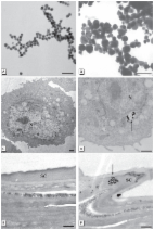
          The use of nanoparticle delivery systems to enhance intracellular penetration of antibiotics and their retention time is becoming popular. The challenge, however, is that the interaction of nanoparticles with biological systems at the cellular level must be established prior to biomedical applications. Ciprofloxacin–cockle shells-derived calcium carbonate (aragonite) nanoparticles (C-CSCCAN) were developed and characterized. Antibacterial activity was determined using a modified disc diffusion protocol on Salmonella Typhimurium ( S. Typhimurium). Biocompatibilittes with macrophage were evaluated using the 3-(4,5-Dimethylthiazol-2-yl)-2,5-diphenyltetrazolium bromide (MTT) and 5-Bromo-2′-deoxyuridine (BrdU) assays. Transcriptional regulation of interleukin 1 beta (IL-1β) was determined using reverse transcriptase-polymerase chain reaction (RT-PCR). C-CSCCAN were spherical in shape, with particle sizes ranging from 11.93 to 22.12 nm. Encapsulation efficiency (EE) and loading content (LC) were 99.5% and 5.9%, respectively, with negative ζ potential. X-ray diffraction patterns revealed strong crystallizations and purity in the formulations. The mean diameter of inhibition zone was 18.6 ± 0.5 mm, which was better than ciprofloxacin alone (11.7 ± 0.9 mm). Study of biocompatability established the cytocompatability of the delivery system without upregulation of IL-1β. The results indicated that ciprofloxacin–nanoparticles enhanced the antibacterial efficacy of the antibiotic, and could act as a suitable delivery system against intracellular infections.

          Related collections

          Most cited references70

          • Record: found
          • Abstract: found
          • Article: not found

          "Nanoantibiotics": a new paradigm for treating infectious diseases using nanomaterials in the antibiotics resistant era.

          Despite the fact that we live in an era of advanced and innovative technologies for elucidating underlying mechanisms of diseases and molecularly designing new drugs, infectious diseases continue to be one of the greatest health challenges worldwide. The main drawbacks for conventional antimicrobial agents are the development of multiple drug resistance and adverse side effects. Drug resistance enforces high dose administration of antibiotics, often generating intolerable toxicity, development of new antibiotics, and requests for significant economic, labor, and time investments. Recently, nontraditional antibiotic agents have been of tremendous interest in overcoming resistance that is developed by several pathogenic microorganisms against most of the commonly used antibiotics. Especially, several classes of antimicrobial nanoparticles (NPs) and nanosized carriers for antibiotics delivery have proven their effectiveness for treating infectious diseases, including antibiotics resistant ones, in vitro as well as in animal models. This review summarizes emerging efforts in combating against infectious diseases, particularly using antimicrobial NPs and antibiotics delivery systems as new tools to tackle the current challenges in treating infectious diseases. Copyright © 2011 Elsevier B.V. All rights reserved.
            Bookmark
            • Record: found
            • Abstract: found
            • Article: not found

            Strain specificity in antimicrobial activity of silver and copper nanoparticles.

            The antimicrobial properties of silver and copper nanoparticles were investigated using Escherichia coli (four strains), Bacillus subtilis and Staphylococcus aureus (three strains). The average sizes of the silver and copper nanoparticles were 3 nm and 9 nm, respectively, as determined through transmission electron microscopy. Energy-dispersive X-ray spectra of silver and copper nanoparticles revealed that while silver was in its pure form, an oxide layer existed on the copper nanoparticles. The bactericidal effect of silver and copper nanoparticles were compared based on diameter of inhibition zone in disk diffusion tests and minimum inhibitory concentration (MIC) and minimum bactericidal concentration (MBC) of nanoparticles dispersed in batch cultures. Bacterial sensitivity to nanoparticles was found to vary depending on the microbial species. Disk diffusion studies with E. coli and S. aureus revealed greater effectiveness of the silver nanoparticles compared to the copper nanoparticles. B. subtilis depicted the highest sensitivity to nanoparticles compared to the other strains and was more adversely affected by the copper nanoparticles. Good correlation was observed between MIC and MBC (r2=0.98) measured in liquid cultures. For copper nanoparticles a good negative correlation was observed between the inhibition zone observed in disk diffusion test and MIC/MBC determined based on liquid cultures with the various strains (r2=-0.75). Although strain-specific variation in MIC/MBC was negligible for S. aureus, some strain-specific variation was observed for E. coli.
              Bookmark
              • Record: found
              • Abstract: found
              • Article: found
              Is Open Access

              Evaluation of Silver Nanoparticle Toxicity in Skin in Vivo and Keratinocytes in Vitro

              Introduction Products using the antimicrobial properties of silver nanoparticles (Ag-nps) may be found in health and consumer products that routinely contact skin. Objectives This study was designed to assess the potential cytotoxicity of Ag-nps in human epidermal keratinocytes (HEKs) and their inflammatory and penetrating potential into porcine skin in vivo. Materials and Methods We used eight different Ag-nps in this study [unwashed/uncoated (20, 50, and 80 nm particle diameter), washed/uncoated (20, 50, and 80 nm), and carbon-coated (25 and 35 nm)]. Skin was dosed topically for 14 consecutive days. HEK viability was assessed by MTT, alamarBlue (aB), and CellTiter 96 AQueous One (96AQ). Release of the proinflammatory mediators interleukin (IL)-1β, IL-6, IL-8, IL-10, and tumor necrosis factor-α (TNF-α) were measured. Results The effect of the unwashed Ag-nps on HEK viability after a 24-hr exposure indicated a significant dose-dependent decrease (p < 0.05) at 0.34 μg/mL with aB and 96AQ and at 1.7 μg/mL with MTT. However, both the washed Ag-nps and carbon-coated Ag-nps showed no significant decrease in viability at any concentration assessed by any of the three assays. For each of the unwashed Ag-nps, we noted a significant increase (p < 0.05) in IL-1β, IL-6, IL-8, and TNF-α concentrations. We observed localization of all Ag-nps in cytoplasmic vacuoles of HEKs. Macroscopic observations showed no gross irritation in porcine skin, whereas microscopic and ultrastructural observations showed areas of focal inflammation and localization of Ag-nps on the surface and in the upper stratum corneum layers of the skin. Conclusion This study provides a better understanding Ag-nps safety in vitro as well as in vivo and a basis for occupational and risk assessment. Ag-nps are nontoxic when dosed in washed Ag-nps solutions or carbon coated.
                Bookmark

                Author and article information

                Contributors
                Role: Academic Editor
                Journal
                Int J Mol Sci
                Int J Mol Sci
                ijms
                International Journal of Molecular Sciences
                MDPI
                1422-0067
                19 May 2016
                May 2016
                : 17
                : 5
                : 713
                Affiliations
                [1 ]Laboratory of Molecular Biomedicine, Institute of Bioscience, Universiti Putra Malaysia, Serdang, Selangor 43400, Malaysia; tjelyakub@ 123456gmail.com (T.I.); mustyimam@ 123456gmail.com (M.U.I.)
                [2 ]Faculty of Food Science and Technology and Laboratory of Natural Product, Institute of Bioscience, Universiti Putra Malaysia, Serdang, Selangor 43400, Malaysia; yaya_rukayadi@ 123456upm.edu.my
                [3 ]Faculty of Veterinary Medicine, Universiti Putra Malaysia, Serdang, Selangor 43400, Malaysia; jajidvm@ 123456yahoo.com (A.Z.J.); naheda_ibrahem@ 123456yahoo.com (N.I.H.); saffanh.jeber@ 123456gmail.com (S.K.M.)
                [4 ]Laboratory of Pharmacology and Toxicology, Faculty of Veterinary Medicine, Universiti Putra Malaysia, Serdang, Selangor 43400, Malaysia; hezmee@ 123456upm.edu.my
                Author notes
                [* ]Correspondence: zuki@ 123456upm.edu.my ; Tel.: +603-89-462-102; Fax: +603-89-472-101
                Article
                ijms-17-00713
                10.3390/ijms17050713
                4881535
                27213349
                aa62ca9d-0035-4d3d-829d-7e24ebea182e
                © 2016 by the authors; licensee MDPI, Basel, Switzerland.

                This article is an open access article distributed under the terms and conditions of the Creative Commons Attribution (CC-BY) license ( http://creativecommons.org/licenses/by/4.0/).

                History
                : 12 January 2016
                : 19 April 2016
                Categories
                Article

                Molecular biology
                antimicrobial resistance,calcium carbonate (aragonite) nanoparticles,ciprofloxacin,intracellular infection,proinflammatory cytokine

                Comments

                Comment on this article