4
views
0
recommends
+1 Recommend
0 collections
    0
    shares
      • Record: found
      • Abstract: found
      • Article: found
      Is Open Access

      Global Proteomics to Study Silica Nanoparticle-Induced Cytotoxicity and Its Mechanisms in HepG2 Cells

      research-article

      Read this article at

      Bookmark
          There is no author summary for this article yet. Authors can add summaries to their articles on ScienceOpen to make them more accessible to a non-specialist audience.

          Abstract

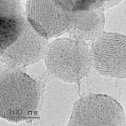
          Silica nanoparticles (SiO 2 NPs) are commonly used in medical and pharmaceutical fields. Research into the cytotoxicity and overall proteomic changes occurring during initial exposure to SiO 2 NPs is limited. We investigated the mechanism of toxicity in human liver cells according to exposure time [0, 4, 10, and 16 h (h)] to SiO 2 NPs through proteomic analysis using mass spectrometry. SiO 2 NP-induced cytotoxicity through various pathways in HepG2 cells. Interestingly, when cells were exposed to SiO 2 NPs for 4 h, the morphology of the cells remained intact, while the expression of proteins involved in mRNA splicing, cell cycle, and mitochondrial function was significantly downregulated. These results show that the toxicity of the nanoparticles affects protein expression even if there is no change in cell morphology at the beginning of exposure to SiO 2 NPs. The levels of reactive oxygen species changed significantly after 10 h of exposure to SiO 2 NPs, and the expression of proteins associated with oxidative phosphorylation, as well as the immune system, was upregulated. Eventually, these changes in protein expression induced HepG2 cell death. This study provides insights into cytotoxicity evaluation at early stages of exposure to SiO 2 NPs through in vitro experiments.

          Related collections

          Most cited references64

          • Record: found
          • Abstract: found
          • Article: found
          Is Open Access

          Principles for characterizing the potential human health effects from exposure to nanomaterials: elements of a screening strategy

          The rapid proliferation of many different engineered nanomaterials (defined as materials designed and produced to have structural features with at least one dimension of 100 nanometers or less) presents a dilemma to regulators regarding hazard identification. The International Life Sciences Institute Research Foundation/Risk Science Institute convened an expert working group to develop a screening strategy for the hazard identification of engineered nanomaterials. The working group report presents the elements of a screening strategy rather than a detailed testing protocol. Based on an evaluation of the limited data currently available, the report presents a broad data gathering strategy applicable to this early stage in the development of a risk assessment process for nanomaterials. Oral, dermal, inhalation, and injection routes of exposure are included recognizing that, depending on use patterns, exposure to nanomaterials may occur by any of these routes. The three key elements of the toxicity screening strategy are: Physicochemical Characteristics, In Vitro Assays (cellular and non-cellular), and In Vivo Assays. There is a strong likelihood that biological activity of nanoparticles will depend on physicochemical parameters not routinely considered in toxicity screening studies. Physicochemical properties that may be important in understanding the toxic effects of test materials include particle size and size distribution, agglomeration state, shape, crystal structure, chemical composition, surface area, surface chemistry, surface charge, and porosity. In vitro techniques allow specific biological and mechanistic pathways to be isolated and tested under controlled conditions, in ways that are not feasible in in vivo tests. Tests are suggested for portal-of-entry toxicity for lungs, skin, and the mucosal membranes, and target organ toxicity for endothelium, blood, spleen, liver, nervous system, heart, and kidney. Non-cellular assessment of nanoparticle durability, protein interactions, complement activation, and pro-oxidant activity is also considered. Tier 1 in vivo assays are proposed for pulmonary, oral, skin and injection exposures, and Tier 2 evaluations for pulmonary exposures are also proposed. Tier 1 evaluations include markers of inflammation, oxidant stress, and cell proliferation in portal-of-entry and selected remote organs and tissues. Tier 2 evaluations for pulmonary exposures could include deposition, translocation, and toxicokinetics and biopersistence studies; effects of multiple exposures; potential effects on the reproductive system, placenta, and fetus; alternative animal models; and mechanistic studies.
            Bookmark
            • Record: found
            • Abstract: found
            • Article: found
            Is Open Access

            Mesoporous silica nanoparticles for drug and gene delivery

            Mesoporous silica nanoparticles (MSNs) are attracting increasing interest for potential biomedical applications. With tailored mesoporous structure, huge surface area and pore volume, selective surface functionality, as well as morphology control, MSNs exhibit high loading capacity for therapeutic agents and controlled release properties if modified with stimuli-responsive groups, polymers or proteins. In this review article, the applications of MSNs in pharmaceutics to improve drug bioavailability, reduce drug toxicity, and deliver with cellular targetability are summarized. Particularly, the exciting progress in the development of MSNs-based effective delivery systems for poorly soluble drugs, anticancer agents, and therapeutic genes are highlighted.
              Bookmark
              • Record: found
              • Abstract: found
              • Article: found
              Is Open Access

              Toxicology of silica nanoparticles: an update

              Large-scale production and use of amorphous silica nanoparticles (SiNPs) have increased the risk of human exposure to SiNPs, while their health effects remain unclear. In this review, scientific papers from 2010 to 2016 were systematically selected and sorted based on in vitro and in vivo studies: to provide an update on SiNPs toxicity and to address the knowledge gaps indicated in the review of Napierska (Part Fibre Toxicol 7:39, 2010). Toxicity of SiNPs in vitro is size, dose, and cell type dependent. SiNPs synthesized by wet route exhibited noticeably different biological effects compared to thermal route-based SiNPs. Amorphous SiNPs (particularly colloidal and stöber) induced toxicity via mechanisms similar to crystalline silica. In vivo, route of administration and physico-chemical properties of SiNPs influences the toxicokinetics. Adverse effects were mainly observed in acutely exposed animals, while no significant signs of toxicity were noted in chronically dosed animals. The correlation between in vitro and in vivo toxicity remains less well established mainly due to improper—unrealistic—dosing both in vitro and in vivo. In conclusion, notwithstanding the multiple studies published in recent years, unambiguous linking of physico-chemical properties of SiNPs types to toxicity, bioavailability, or human health effects is not yet possible.
                Bookmark

                Author and article information

                Contributors
                Role: Academic Editor
                Journal
                Biomolecules
                Biomolecules
                biomolecules
                Biomolecules
                MDPI
                2218-273X
                02 March 2021
                March 2021
                : 11
                : 3
                : 375
                Affiliations
                [1 ]Bioimaging Team, Safety Measurement Institute, Korea Research Institute of Standards and Science (KRISS), Daejeon 34113, Korea; sylee12@ 123456kriss.re.kr
                [2 ]Nano-Safety Team, Safety Measurement Institute, Korea Research Institute of Standards and Science (KRISS), Daejeon 34113, Korea; inyoungkim@ 123456kriss.re.kr (I.Y.K.); mbheo@ 123456kriss.re.kr (M.B.H.)
                [3 ]Disease Target Structure Research Center, Korea Research Institute of Bioscience and Biotechnology (KRIBB), Daejeon 34141, Korea; jhdal@ 123456kribb.re.kr
                Author notes
                [* ]Correspondence: yeskyoung@ 123456kriss.re.kr (J.G.S.); tglee@ 123456kriss.re.kr (T.G.L.); Tel.: +82-42-868-5751 (J.G.S.); +82-42-868-5003 (T.G.L.)
                Author information
                https://orcid.org/0000-0002-6568-9377
                Article
                biomolecules-11-00375
                10.3390/biom11030375
                8000044
                33801561
                aaa16b78-9be6-40b5-b9da-7eff33dec636
                © 2021 by the authors.

                Licensee MDPI, Basel, Switzerland. This article is an open access article distributed under the terms and conditions of the Creative Commons Attribution (CC BY) license ( http://creativecommons.org/licenses/by/4.0/).

                History
                : 20 January 2021
                : 24 February 2021
                Categories
                Article

                silica nanoparticles,cytotoxicity,label-free quantification,proteomic analysis,mass spectrometry

                Comments

                Comment on this article