34
views
0
recommends
+1 Recommend
0 collections
    0
    shares
      • Record: found
      • Abstract: found
      • Article: found
      Is Open Access

      Effect of nano-hydroxyapatite coating on the osteoinductivity of porous biphasic calcium phosphate ceramics

      research-article

      Read this article at

      Bookmark
          There is no author summary for this article yet. Authors can add summaries to their articles on ScienceOpen to make them more accessible to a non-specialist audience.

          Abstract

          Background

          Porous biphasic calcium phosphate (BCP) ceramics exhibit good biocompatibility and bone conduction but are not inherently osteoinductive. To overcome this disadvantage, we coated conventional porous BCP ceramics with nano-hydroxyapatite (nHA). nHA was chosen as a coating material due to its high osteoinductive potential.

          Methods

          We used a hydrothermal deposition method to coat conventional porous BCP ceramics with nHA and assessed the effects of the coating on the physical and mechanical properties of the underlying BCP. Next, its effects on mesenchymal stem cell (MSC) attachment, proliferation, viability, and osteogenic differentiation were investigated.

          Results

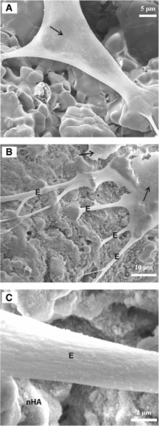
          nHA formed a deposited layer on the BCP surface, and synthesized nHA had a rod-like shape with lengths ranging from ~50–200 nm and diameters from ~15–30 mm. The nHA coating did not significantly affect the density, porosity, flexural strength, or compressive strength of the underlying BCP ( P > 0.1). Scanning electron microscopy showed MSC attachment to the scaffolds, with a healthy morphology and anchorage to nHA crystals via cytoplasmic processes. The densities of MSCs attached on BCP and nHA-coated BCP scaffolds were 62 ± 26 cells/mm 2 and 63 ± 27 cells/mm 2 ( P > 0.1), respectively, after 1 day and 415 ± 62 cells/mm 2 and 541 ± 35 cells/mm 2 ( P < 0.05) respectively, after 14 days. According to an MTT assay, MSC viability was higher on nHA-coated BCP scaffolds than on BCP scaffolds ( P < 0.05). In addition, MSCs on nHA-coated BCP scaffolds produced more alkaline phosphatase, collagen type I, and osteocalcin than MSCs on BCP scaffolds ( P < 0.05).

          Conclusions

          Our results demonstrate that BCP scaffolds coated with nHA were more conducive for MSC adhesion, proliferation, and osteogenic differentiation than conventional, uncoated BCP scaffolds, indicating that nHA coating can enhance the osteoinductive potential of BCP ceramics, making this material more suitable for applications in bone tissue engineering.

          Related collections

          Most cited references33

          • Record: found
          • Abstract: not found
          • Article: not found

          Nature’s hierarchical materials

            Bookmark
            • Record: found
            • Abstract: found
            • Article: not found

            Enhanced functions of osteoblasts on nanophase ceramics.

            T. Webster (2000)
            Select functions of osteoblasts (bone-forming cells) on nanophase (materials with grain sizes less than 100 nm) alumina, titania, and hydroxyapatite (HA) were investigated using in vitro cellular models. Compared to conventional ceramics, surface occupancy of osteoblast colonies was significantly less on all nanophase ceramics tested in the present study after 4 and 6 days of culture. Osteoblast proliferation was significantly greater on nanophase alumina, titania, and HA than on conventional formulations of the same ceramic after 3 and 5 days. More importantly, compared to conventional ceramics, synthesis of alkaline phosphatase and deposition of calcium-containing mineral was significantly greater by osteoblasts cultured on nanophase than on conventional ceramics after 21 and 28 days. The results of the present study provided the first evidence of enhanced long-term (on the order of days to weeks) functions of osteoblasts cultured on nanophase ceramics; in this manner, nanophase ceramics clearly represent a unique and promising class of orthopaedic/dental implant formulations with improved osseointegrative properties.
              Bookmark
              • Record: found
              • Abstract: found
              • Article: not found

              The effect of calcium ion concentration on osteoblast viability, proliferation and differentiation in monolayer and 3D culture.

              Our research group aims to develop an osteochondral composite using type II collagen gel with hydroxyapatite (HAp) deposited on one side. Soaking gels in Ca2+ and phosphate solution is indispensable to HAp deposition, so relationships between cell behavior and Ca2+ concentration were examined in two- and three-dimensional cultures. The present results indicate that 2-4 mM Ca2+ is suitable for proliferation and survival of osteoblasts, whereas slightly higher concentrations (6-8 mM) favor osteoblast differentiation and matrix mineralization in both 2- and 3-dimensional cultures. Higher concentrations (>10 mM) are cytotoxic. Purely from the perspective of calcium deposition, higher concentrations lead to increased accumulation of Ca2+. Culturing cells in phosphate-containing gel in media with Ca2+ also leads to time-dependent formation of HAp in the gel. Considering the viability of embedded cells, culturing scaffolds in media with Ca2+ concentrations around 5mM is useful for both HAp deposition and osteoblast behavior.
                Bookmark

                Author and article information

                Contributors
                Journal
                BMC Musculoskelet Disord
                BMC Musculoskelet Disord
                BMC Musculoskeletal Disorders
                BioMed Central
                1471-2474
                2014
                1 April 2014
                : 15
                : 114
                Affiliations
                [1 ]Department of Spine Surgery, Xiangya Hospital, Central South University, Changsha, 410008, China
                [2 ]Center for Medical Experiments, Third Xiangya Hospital, Central South University, Changsha, 410013, China
                [3 ]Department of Sports Medicine, Research Center of Sports Medicine, Xiangya Hospital, Central South University, Changsha, 410008, China
                Article
                1471-2474-15-114
                10.1186/1471-2474-15-114
                3994218
                24690170
                abe48e40-c17b-42d8-ad83-5f4c176e5df2
                Copyright © 2014 Hu et al.; licensee BioMed Central Ltd.

                This is an Open Access article distributed under the terms of the Creative Commons Attribution License ( http://creativecommons.org/licenses/by/2.0), which permits unrestricted use, distribution, and reproduction in any medium, provided the original work is properly credited. The Creative Commons Public Domain Dedication waiver ( http://creativecommons.org/publicdomain/zero/1.0/) applies to the data made available in this article, unless otherwise stated.

                History
                : 2 October 2013
                : 5 March 2014
                Categories
                Research Article

                Orthopedics
                mesenchymal stem cell,cell proliferation,osteogenesis,bone regeneration,biocompatibility,nanotopography

                Comments

                Comment on this article