21
views
0
recommends
+1 Recommend
0 collections
    0
    shares
      • Record: found
      • Abstract: found
      • Article: found
      Is Open Access

      Epithelial Intermediate Filaments: Guardians against Microbial Infection?

      review-article
      * , *
      Cells
      MDPI
      epithelium, keratin, barrier, pathogen, virus, bacterium, parasite, Caenorhabditis elegans

      Read this article at

      Bookmark
          There is no author summary for this article yet. Authors can add summaries to their articles on ScienceOpen to make them more accessible to a non-specialist audience.

          Abstract

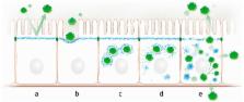
          Intermediate filaments are abundant cytoskeletal components of epithelial tissues. They have been implicated in overall stress protection. A hitherto poorly investigated area of research is the function of intermediate filaments as a barrier to microbial infection. This review summarizes the accumulating knowledge about this interaction. It first emphasizes the unique spatial organization of the keratin intermediate filament cytoskeleton in different epithelial tissues to protect the organism against microbial insults. We then present examples of direct interaction between viral, bacterial, and parasitic proteins and the intermediate filament system and describe how this affects the microbe-host interaction by modulating the epithelial cytoskeleton, the progression of infection, and host response. These observations not only provide novel insights into the dynamics and function of intermediate filaments but also indicate future avenues to combat microbial infection.

          Related collections

          Most cited references153

          • Record: found
          • Abstract: found
          • Article: not found

          Diarrheagenic Escherichia coli.

          Escherichia coli is the predominant nonpathogenic facultative flora of the human intestine. Some E. coli strains, however, have developed the ability to cause disease of the gastrointestinal, urinary, or central nervous system in even the most robust human hosts. Diarrheagenic strains of E. coli can be divided into at least six different categories with corresponding distinct pathogenic schemes. Taken together, these organisms probably represent the most common cause of pediatric diarrhea worldwide. Several distinct clinical syndromes accompany infection with diarrheagenic E. coli categories, including traveler's diarrhea (enterotoxigenic E. coli), hemorrhagic colitis and hemolytic-uremic syndrome (enterohemorrhagic E. coli), persistent diarrhea (enteroaggregative E. coli), and watery diarrhea of infants (entero-pathogenic E. coli). This review discusses the current level of understanding of the pathogenesis of the diarrheagenic E. coli strains and describes how their pathogenic schemes underlie the clinical manifestations, diagnostic approach, and epidemiologic investigation of these important pathogens.
            Bookmark
            • Record: found
            • Abstract: found
            • Article: not found

            Increased expression of interleukin 17 in inflammatory bowel disease.

            Interleukin (IL) 17 is a cytokine which exerts strong proinflammatory activities. In this study we evaluated changes in IL-17 expression in the inflamed mucosa and in the serum of patients with inflammatory bowel disease (IBD). Tissue samples were obtained endoscopically or surgically from patients with ulcerative colitis (UC) (n=20), Crohn's disease (CD) (n=20), infectious colitis (n=5), ischaemic colitis (n=8), and normal colorectal tissues (n=15). IL-17 expression was evaluated by a standard immunohistochemical procedure. Serum IL-17 levels were determined by ELISA. IL-17 mRNA expression was analysed by reverse transcriptase-polymerase chain reaction. IL-17 expression was not detected in samples from normal colonic mucosa, infectious colitis, or ischaemic colitis. In the inflamed mucosa of active UC and CD patients, IL-17 expression was clearly detectable in CD3(+) T cells or CD68(+) monocytes/macrophages. The average number of IL-17(+) cells was significantly increased in active UC and CD patients compared with inactive patients. IL-17 mRNA expression was not detected in normal mucosa but was detectable in the mucosa from active UC and CD patients. IL-17 was not detected in the sera from normal individuals, infectious colitis, or ischaemic colitis patients but IL-17 levels were significantly elevated in IBD patients. IL-17 expression in the mucosa and serum was increased in IBD patients. It is likely that IL-17 expression in IBD may be associated with altered immune and inflammatory responses in the intestinal mucosa.
              Bookmark
              • Record: found
              • Abstract: found
              • Article: not found

              Structure and functions of keratin proteins in simple, stratified, keratinized and cornified epithelia.

              Historically, the term 'keratin' stood for all of the proteins extracted from skin modifications, such as horns, claws and hooves. Subsequently, it was realized that this keratin is actually a mixture of keratins, keratin filament-associated proteins and other proteins, such as enzymes. Keratins were then defined as certain filament-forming proteins with specific physicochemical properties and extracted from the cornified layer of the epidermis, whereas those filament-forming proteins that were extracted from the living layers of the epidermis were grouped as 'prekeratins' or 'cytokeratins'. Currently, the term 'keratin' covers all intermediate filament-forming proteins with specific physicochemical properties and produced in any vertebrate epithelia. Similarly, the nomenclature of epithelia as cornified, keratinized or non-keratinized is based historically on the notion that only the epidermis of skin modifications such as horns, claws and hooves is cornified, that the non-modified epidermis is a keratinized stratified epithelium, and that all other stratified and non-stratified epithelia are non-keratinized epithelia. At this point in time, the concepts of keratins and of keratinized or cornified epithelia need clarification and revision concerning the structure and function of keratin and keratin filaments in various epithelia of different species, as well as of keratin genes and their modifications, in view of recent research, such as the sequencing of keratin proteins and their genes, cell culture, transfection of epithelial cells, immunohistochemistry and immunoblotting. Recently, new functions of keratins and keratin filaments in cell signaling and intracellular vesicle transport have been discovered. It is currently understood that all stratified epithelia are keratinized and that some of these keratinized stratified epithelia cornify by forming a Stratum corneum. The processes of keratinization and cornification in skin modifications are different especially with respect to the keratins that are produced. Future research in keratins will provide a better understanding of the processes of keratinization and cornification of stratified epithelia, including those of skin modifications, of the adaptability of epithelia in general, of skin diseases, and of the changes in structure and function of epithelia in the course of evolution. This review focuses on keratins and keratin filaments in mammalian tissue but keratins in the tissues of some other vertebrates are also considered.
                Bookmark

                Author and article information

                Contributors
                Role: Academic Editor
                Journal
                Cells
                Cells
                cells
                Cells
                MDPI
                2073-4409
                27 June 2016
                September 2016
                : 5
                : 3
                : 29
                Affiliations
                Institute of Molecular and Cellular Anatomy, RWTH Aachen University, Wendlingweg 2, 52074 Aachen, Germany
                Author notes
                [* ]Correspondence: fgeisler@ 123456ukaachen.de (F.G.); rleube@ 123456ukaachen.de (R.E.L.); Tel.: +49-241-80-89107 (R.E.L.); +49-241-80-88927 (F.G.); Fax: +49-241-80-82508 (F.G. and R.E.L.)
                Article
                cells-05-00029
                10.3390/cells5030029
                5040971
                27355965
                ade2ac13-6d10-48ad-9820-64b67034448b
                © 2016 by the authors; licensee MDPI, Basel, Switzerland.

                This article is an open access article distributed under the terms and conditions of the Creative Commons Attribution (CC-BY) license ( http://creativecommons.org/licenses/by/4.0/).

                History
                : 11 May 2016
                : 21 June 2016
                Categories
                Review

                epithelium,keratin,barrier,pathogen,virus,bacterium,parasite,caenorhabditis elegans

                Comments

                Comment on this article