17
views
0
recommends
+1 Recommend
0 collections
    0
    shares
      • Record: found
      • Abstract: found
      • Article: found
      Is Open Access

      Pattern Recognition Approaches for Breast Cancer DCE-MRI Classification: A Systematic Review

      review-article

      Read this article at

      Bookmark
          There is no author summary for this article yet. Authors can add summaries to their articles on ScienceOpen to make them more accessible to a non-specialist audience.

          Abstract

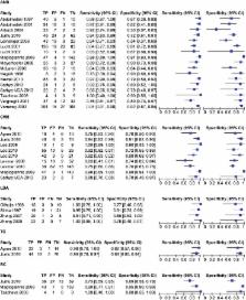
          We performed a systematic review of several pattern analysis approaches for classifying breast lesions using dynamic, morphological, and textural features in dynamic contrast-enhanced magnetic resonance imaging (DCE-MRI). Several machine learning approaches, namely artificial neural networks (ANN), support vector machines (SVM), linear discriminant analysis (LDA), tree-based classifiers (TC), and Bayesian classifiers (BC), and features used for classification are described. The findings of a systematic review of 26 studies are presented. The sensitivity and specificity are respectively 91 and 83 % for ANN, 85 and 82 % for SVM, 96 and 85 % for LDA, 92 and 87 % for TC, and 82 and 85 % for BC. The sensitivity and specificity are respectively 82 and 74 % for dynamic features, 93 and 60 % for morphological features, 88 and 81 % for textural features, 95 and 86 % for a combination of dynamic and morphological features, and 88 and 84 % for a combination of dynamic, morphological, and other features. LDA and TC have the best performance. A combination of dynamic and morphological features gives the best performance.

          Related collections

          Most cited references40

          • Record: found
          • Abstract: found
          • Article: not found

          Dynamic breast MR imaging: are signal intensity time course data useful for differential diagnosis of enhancing lesions?

          To assess the relevance of the signal intensity time course for the differential diagnosis of enhancing lesions in dynamic magnetic resonance (MR) imaging of the breast. Two hundred sixty-six breast lesions were examined with a two-dimensional dynamic MR imaging series and subtraction postprocessing. Time-signal intensity curves of the lesions were obtained and classified according to their shapes as type I, which was steady enhancement; type II, plateau of signal intensity; or type III, washout of signal intensity. Enhancement rates and curve types of benign and malignant lesions were compared. There were 101 malignant and 165 benign lesions. The distribution of curve types for breast cancers was type I, 8.9%; type II, 33.6%; and type III, 57.4%. The distribution of curve types for benign lesions was type I, 83.0%; type II, 11.5%; and type III, 5.5%. The distributions proved significantly different (chi 2 = 139.6; P < .001). The diagnostic indices for signal intensity time course were sensitivity, 91%; specificity, 83%; and diagnostic accuracy, 86%. The diagnostic indices for the enhancement rate were sensitivity, 91%; specificity, 37%; and diagnostic accuracy, 58%. The shape of the time-signal intensity curve is an important criterion in differentiating benign and malignant enhancing lesions in dynamic breast MR imaging. A type III time course is a strong indicator of malignancy and is independent of other criteria.
            Bookmark
            • Record: found
            • Abstract: not found
            • Book: not found

            Introduction to Statistical Pattern Recognition

              Bookmark
              • Record: found
              • Abstract: found
              • Article: not found

              Quantitative analysis of lesion morphology and texture features for diagnostic prediction in breast MRI.

              To investigate the feasibility using quantitative morphology/texture features of breast lesions for diagnostic prediction, and to explore the association of computerized features with lesion phenotype appearance on magnetic resonance imaging. Forty-three malignant/28 benign lesions were used in this study. A systematic approach from automated lesion segmentation, quantitative feature extraction, diagnostic feature selection using an artificial neural network (ANN), and lesion classification was carried out. Eight morphologic parameters and 10 gray level co-occurrence matrix texture features were obtained from each lesion. The diagnostic performance of selected features to differentiate between malignant and benign lesions was analyzed using receiver-operating characteristic analysis. Six features were selected by an ANN using leave-one-out cross validation, including compactness, normalized radial length entropy, volume, gray level entropy, gray level sum average, and homogeneity. The area under the receiver-operating characteristic curve was 0.86. When dividing the database into half training and half validation set, a classifier of five features selected in the half training set achieved an area under the curve of 0.82 in the other half validation set. The selected morphology feature "compactness" was associated with shape and margin in the Breast Imaging Reporting and Data System lexicon, round shape and smooth margin for the benign lesions, and more irregular shape for the malignant lesions. The selected texture features were associated with homogeneous/heterogeneous patterns and the enhancement intensity. The malignant lesions had higher intensity and broader distribution on the enhancement histogram (more heterogeneous) compared to the benign lesions. Quantitative analysis of morphology/texture features of breast lesions was feasible, and these features could be selected by an ANN to form a classifier for differential diagnosis. Establishing the link between computer-based features and visual descriptors defined in the BI-RADS lexicon will provide the foundation for the acceptance of quantitative diagnostic features in the development of computer-aided diagnosis.
                Bookmark

                Author and article information

                Contributors
                00390815903738 , r.fusco@istitutotumori.na.it
                Journal
                J Med Biol Eng
                J Med Biol Eng
                Journal of Medical and Biological Engineering
                Springer Berlin Heidelberg (Berlin/Heidelberg )
                1609-0985
                31 August 2016
                31 August 2016
                2016
                : 36
                : 4
                : 449-459
                Affiliations
                [1 ]Department of Diagnostic Imaging, metabolic and radiant Therapy, National Cancer Institute of Naples “Pascale Foundation”, Via Mariano Semmola 80131, Naples, Italy
                [2 ]Department of Electrical Engineering and Information Technologies, University ‘Federico II’, Via Claudio 80125, Naples, Italy
                Article
                163
                10.1007/s40846-016-0163-7
                5016558
                ae2dc404-340c-4ffa-88ef-05dce7b175d0
                © The Author(s) 2016

                Open AccessThis article is distributed under the terms of the Creative Commons Attribution 4.0 International License ( http://creativecommons.org/licenses/by/4.0/), which permits unrestricted use, distribution, and reproduction in any medium, provided you give appropriate credit to the original author(s) and the source, provide a link to the Creative Commons license, and indicate if changes were made.

                History
                : 30 September 2015
                : 29 March 2016
                Categories
                Review Article
                Custom metadata
                © Taiwanese Society of Biomedical Engineering 2016

                dynamic contrast-enhanced magnetic resonance imaging (dce-mri),breast cancer,patter recognition approach,classification

                Comments

                Comment on this article