34
views
0
recommends
+1 Recommend
0 collections
    0
    shares
      • Record: found
      • Abstract: found
      • Article: found
      Is Open Access

      Exploring the Neural Basis of Real-Life Joint Action: Measuring Brain Activation during Joint Table Setting with Functional Near-Infrared Spectroscopy

      research-article

      Read this article at

      Bookmark
          There is no author summary for this article yet. Authors can add summaries to their articles on ScienceOpen to make them more accessible to a non-specialist audience.

          Abstract

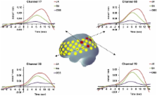
          Many every-day life situations require two or more individuals to execute actions together. Assessing brain activation during naturalistic tasks to uncover relevant processes underlying such real-life joint action situations has remained a methodological challenge. In the present study, we introduce a novel joint action paradigm that enables the assessment of brain activation during real-life joint action tasks using functional near-infrared spectroscopy (fNIRS). We monitored brain activation of participants who coordinated complex actions with a partner sitting opposite them. Participants performed table setting tasks, either alone (solo action) or in cooperation with a partner (joint action), or they observed the partner performing the task (action observation). Comparing joint action and solo action revealed stronger activation (higher [oxy-Hb]-concentration) during joint action in a number of areas. Among these were areas in the inferior parietal lobule (IPL) that additionally showed an overlap of activation during action observation and solo action. Areas with such a close link between action observation and action execution have been associated with action simulation processes. The magnitude of activation in these IPL areas also varied according to joint action type and its respective demand on action simulation. The results validate fNIRS as an imaging technique for exploring the functional correlates of interindividual action coordination in real-life settings and suggest that coordinating actions in real-life situations requires simulating the actions of the partner.

          Related collections

          Most cited references34

          • Record: found
          • Abstract: found
          • Article: not found

          Mirror neurons and the simulation theory of mind-reading.

          V Gallese (1998)
          A new class of visuomotor neuron has been recently discovered in the monkey's premotor cortex: mirror neurons. These neurons respond both when a particular action is performed by the recorded monkey and when the same action, performed by another individual, is observed. Mirror neurons appear to form a cortical system matching observation and execution of goal-related motor actions. Experimental evidence suggests that a similar matching system also exists in humans. What might be the functional role of this matching system? One possible function is to enable an organism to detect certain mental states of observed conspecifics. This function might be part of, or a precursor to, a more general mind-reading ability. Two different accounts of mind-reading have been suggested. According to `theory theory', mental states are represented as inferred posits of a naive theory. According to `simulation theory', other people's mental states are represented by adopting their perspective: by tracking or matching their states with resonant states of one's own. The activity of mirror neurons, and the fact that observers undergo motor facilitation in the same muscular groups as those utilized by target agents, are findings that accord well with simulation theory but would not be predicted by theory theory.
            Bookmark
            • Record: found
            • Abstract: found
            • Article: not found

            Automatic 3D intersubject registration of MR volumetric data in standardized Talairach space.

            In both diagnostic and research applications, the interpretation of MR images of the human brain is facilitated when different data sets can be compared by visual inspection of equivalent anatomical planes. Quantitative analysis with predefined atlas templates often requires the initial alignment of atlas and image planes. Unfortunately, the axial planes acquired during separate scanning sessions are often different in their relative position and orientation, and these slices are not coplanar with those in the atlas. We have developed a completely automatic method to register a given volumetric data set with Talairach stereotaxic coordinate system. The registration method is based on multi-scale, three-dimensional (3D) cross-correlation with an average (n > 300) MR brain image volume aligned with the Talariach stereotaxic space. Once the data set is re-sampled by the transformation recovered by the algorithm, atlas slices can be directly superimposed on the corresponding slices of the re-sampled volume. the use of such a standardized space also allows the direct comparison, voxel to voxel, of two or more data sets brought into stereotaxic space. With use of a two-tailed Student t test for paired samples, there was no significant difference in the transformation parameters recovered by the automatic algorithm when compared with two manual landmark-based methods (p > 0.1 for all parameters except y-scale, where p > 0.05). Using root-mean-square difference between normalized voxel intensities as an unbiased measure of registration, we show that when estimated and averaged over 60 volumetric MR images in standard space, this measure was 30% lower for the automatic technique than the manual method, indicating better registrations. Likewise, the automatic method showed a 57% reduction in standard deviation, implying a more stable technique. The algorithm is able to recover the transformation even when data are missing from the top or bottom of the volume. We present a fully automatic registration method to map volumetric data into stereotaxic space that yields results comparable with those of manually based techniques. The method requires no manual identification of points or contours and therefore does not suffer the drawbacks involved in user intervention such as reproducibility and interobserver variability.
              Bookmark
              • Record: found
              • Abstract: found
              • Article: not found

              Three-dimensional probabilistic anatomical cranio-cerebral correlation via the international 10-20 system oriented for transcranial functional brain mapping.

              The recent advent of multichannel near-infrared spectroscopy (NIRS) has expanded its technical potential for human brain mapping. However, NIRS measurement has a technical drawback in that it measures cortical activities from the head surface without anatomical information of the object to be measured. This problem is also found in transcranial magnetic stimulation (TMS) that transcranially activates or inactivates the cortical surface. To overcome this drawback, we examined cranio-cerebral correlation using magnetic resonance imaging (MRI) via the guidance of the international 10-20 system for electrode placement, which had originally been developed for electroencephalography. We projected the 10-20 standard cranial positions over the cerebral cortical surface. After examining the cranio-cerebral correspondence for 17 healthy adults, we normalized the 10-20 cortical projection points of the subjects to the standard Montreal Neurological Institute (MNI) and Talairach stereotactic coordinates and obtained their probabilistic distributions. We also expressed the anatomical structures for the 10-20 cortical projection points probabilistically. Next, we examined the distance between the cortical surface and the head surface along the scalp and created a cortical surface depth map. We found that the locations of 10-20 cortical projection points in the standard MNI or Talairach space could be estimated with an average standard deviation of 8 mm. This study provided an initial step toward establishing a three-dimensional probabilistic anatomical platform that enables intra- and intermodal comparisons of NIRS and TMS brain imaging data.
                Bookmark

                Author and article information

                Journal
                Front Hum Neurosci
                Front. Hum. Neurosci.
                Frontiers in Human Neuroscience
                Frontiers Research Foundation
                1662-5161
                08 September 2011
                2011
                : 5
                : 95
                Affiliations
                [1] 1simpleCenter of Excellence ‘Cognitive Interaction Technology’, and Clinical Linguistics, Bielefeld University Bielefeld, Germany
                [2] 2simpleBerlin School of Mind and Brain, Humboldt-Universität zu Berlin Berlin, Germany
                [3] 3simpleDepartment of Psychiatry and Psychotherapy, University of Tübingen Tübingen, Germany
                [4] 4simpleDepartment of Psychiatry, Psychosomatics and Psychotherapy, University of Würzburg Würzburg, Germany
                Author notes

                Edited by: Shuhei Yamaguchi, Shimane University, Japan

                Reviewed by: Hidenao Fukuyama, Kyoto University, Japan; Eiju Watanabe, Jichi Medical University, Japan

                *Correspondence: Johanna Egetemeir, Center of Excellence ‘Cognitive Interaction Technology’, Bielefeld University, Universitätsstr. 25, 33615 Bielefeld, Germany. e-mail: johanna.egetemeir@ 123456uni-bielefeld.de
                Article
                10.3389/fnhum.2011.00095
                3168792
                21927603
                aea3ece5-12b6-421f-a410-a4d8c0faad9e
                Copyright © 2011 Egetemeir, Stenneken, Koehler, Fallgatter and Herrmann.

                This is an open-access article subject to a non-exclusive license between the authors and Frontiers Media SA, which permits use, distribution and reproduction in other forums, provided the original authors and source are credited and other Frontiers conditions are complied with.

                History
                : 14 June 2011
                : 17 August 2011
                Page count
                Figures: 4, Tables: 1, Equations: 0, References: 41, Pages: 9, Words: 7774
                Categories
                Neuroscience
                Original Research

                Neurosciences
                neuroimaging,joint action,social interaction,real-life interaction,simulation,fnirs
                Neurosciences
                neuroimaging, joint action, social interaction, real-life interaction, simulation, fnirs

                Comments

                Comment on this article