70
views
0
recommends
+1 Recommend
0 collections
    0
    shares
      • Record: found
      • Abstract: found
      • Article: found
      Is Open Access

      Comparison of carotid and lower limb atherosclerotic lesions in both previously known and newly diagnosed type 2 diabetes mellitus

      research-article

      Read this article at

      Bookmark
          There is no author summary for this article yet. Authors can add summaries to their articles on ScienceOpen to make them more accessible to a non-specialist audience.

          Abstract

          Aims/Introduction

          To compare carotid and lower limb atherosclerotic lesions, and examine if carotid atherosclerotic lesions are in line with lower limb atherosclerotic lesions, and can reflect generalized atherosclerosis in inpatients with type 2 diabetes.

          Materials and Methods

          This was an observational study carried out in 867 Chinese inpatients with type 2 diabetes, including 573 previously known and 294 newly diagnosed patients. Ultrasonographic assessments of intima-media thickness (IMT), plaques, and stenosis in the carotid and lower limb arteries were evaluated. Atherosclerotic lesions between the carotid and lower limb arteries were compared in both previously known and newly diagnosed diabetes, respectively.

          Results

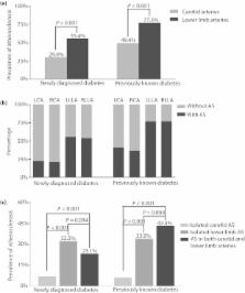
          In both the known (77.3% vs 49.4%, P < 0.001) and the newly diagnosed diabetes (55.4% vs 29.9%, P < 0.001), the prevalence of atherosclerotic plaques was significantly higher in the lower limb arteries than in the carotid arteries. Likewise, the prevalence of stenosis was also significantly higher ( P < 0.001) in the lower limb arteries (16.9%) than in the carotid arteries (4.2%) in the established diabetes patients. However, there was no significant difference in the mean IMT between common carotid and common femoral arteries in both the previously known (0.90 ± 0.24 mm vs 0.89 ± 0.20 mm, P = 0.675) and the newly diagnosed diabetes patients (0.86 ± 0.22 mm vs 0.85 ± 0.16 mm, P = 0.436).

          Conclusions

          Carotid plaques might underestimate generalized plaques in inpatients with type 2 diabetes, as shown by its significantly lower prevalence compared with that of the lower extremity arteries. A combined carotid and lower limb ultrasound examination can improve the detection of atherosclerotic lesions in inpatients with type 2 diabetes.

          Related collections

          Most cited references31

          • Record: found
          • Abstract: found
          • Article: not found

          Carotid artery stenosis: gray-scale and Doppler US diagnosis--Society of Radiologists in Ultrasound Consensus Conference.

          The Society of Radiologists in Ultrasound convened a multidisciplinary panel of experts in the field of vascular ultrasonography (US) to come to a consensus regarding Doppler US for assistance in the diagnosis of carotid artery stenosis. The panel's consensus statement is believed to represent a reasonable position on the basis of analysis of available literature and panelists' experience. Key elements of the statement include the following: (a) All internal carotid artery (ICA) examinations should be performed with gray-scale, color Doppler, and spectral Doppler US. (b) The degree of stenosis determined at gray-scale and Doppler US should be stratified into the categories of normal (no stenosis), or =70% stenosis to near occlusion, near occlusion, and total occlusion. (c) ICA peak systolic velocity (PSV) and presence of plaque on gray-scale and/or color Doppler images are primarily used in diagnosis and grading of ICA stenosis; two additional parameters, ICA-to-common carotid artery PSV ratio and ICA end-diastolic velocity may also be used when clinical or technical factors raise concern that ICA PSV may not be representative of the extent of disease. (d) ICA should be diagnosed as (i) normal when ICA PSV is less than 125 cm/sec and no plaque or intimal thickening is visible; (ii) or =70% stenosis to near occlusion when ICA PSV is greater than 230 cm/sec and visible plaque and lumen narrowing are seen; (v) near occlusion when there is a markedly narrowed lumen at color Doppler US; and (vi) total occlusion when there is no detectable patent lumen at gray-scale US and no flow at spectral, power, and color Doppler US. (e) The final report should discuss velocity measurements and gray-scale and color Doppler findings. Study limitations should be noted when they exist. The conclusion should state an estimated degree of ICA stenosis as reflected in the above categories. The panel also considered various technical aspects of carotid US and methods for quality assessment and identified several important unanswered questions meriting future research. Copyright RSNA, 2003
            Bookmark
            • Record: found
            • Abstract: found
            • Article: found

            Mannheim Intima-Media Thickness Consensus

            Intima-media thickness (IMT) is increasingly used in clinical trials as a surrogate end point for determining the success of interventions that lower risk factors for atherosclerosis. The necessity for unified criteria to distinguish early atherosclerotic plaque formation from thickening of IMT and to standardize IMT measurements is addressed in this consensus statement. Plaque is defined as a focal structure that encroaches into the arterial lumen of at least 0.5 mm or 50% of the surrounding IMT value or demonstrates a thickness of ≧1.5 mm as measured from the media-adventitia interface to the intima-lumen interface. Standard use of IMT measurements is recommended in all epidemiological and interventional trials dealing with vascular diseases to improve characterization of the population investigated. The consensus concludes that there is no need to ‘treat IMT values’ nor to monitor IMT values in individual patients apart from few exceptions. Although IMT has been suggested to represent an important risk marker, it does not fulfill the characteristics of an accepted risk factor. Standardized methods recommended in this consensus statement will foster homogenous data collection and analysis. This will help to improve the power of studies incorporating IMT measurements and to facilitate the merging of large databases for meta-analyses.
              Bookmark
              • Record: found
              • Abstract: found
              • Article: not found

              Carotid plaque, intima media thickness, cardiovascular risk factors, and prevalent cardiovascular disease in men and women: the British Regional Heart Study.

              B-mode ultrasound is a noninvasive method of examining the walls of peripheral arteries and provides measures of the intima-media thickness (IMT) at various sites (common carotid artery, bifurcation, internal carotid artery) and of plaques that may indicate early presymptomatic disease. The reported associations between cardiovascular risk factors, clinical disease, IMT, and plaques are inconsistent. We sought to clarify these relationships in a large, representative sample of men and women living in 2 British towns. The study was performed during 1996 in 2 towns (Dewsbury and Maidstone) of the British Regional Heart Study that have an approximately 2-fold difference in coronary heart disease risk. The male participants were drawn from the British Regional Heart Study and were recruited in 1978-1980 and form part of a national cohort study of 7735 men. A random sample of women of similar age to the men (55 to 77 years) was also selected from the age-sex register of the general practices used in the original survey. A wide range of data on social, lifestyle, and physiological factors, cardiovascular disease symptoms, and diagnoses was collected. Measures of right and left common carotid IMT (IMTcca) and bifurcation IMT (IMTbif) were made, and the arteries were examined for plaques 1.5 cm above and below the flow divider. Totals of 425 men and 375 women were surveyed (mean age, 66 years; range, 56 to 77 years). The mean (SD) IMTcca observed were 0. 84 (0.21) and 0.75 (0.16) mm for men and women, respectively. The mean (SD) IMTbif were 1.69 (0.61) and 1.50 (0.77) mm for men and women, respectively. The correlation between IMTcca and IMTbif was similar in men (r=0.36) and women (r=0.38). There were no differences in mean IMTcca or IMTbif between the 2 towns. Carotid plaques were very common, affecting 57% (n=239) of men and 58% (n=211) of women. Severe carotid plaques with flow disturbance were rare, affecting 9 men (2%) and 6 women (1.6%). Plaques increased in prevalence with age, affecting 49% men and 39% of women aged 70 years. Plaques were most common among men in Dewsbury (79% affected) and least common among men in Maidstone (34% affected). IMTcca showed a different pattern of association with cardiovascular risk factors from IMTbif and was associated with age, SBP, and FEV1 but not with social, lifestyle, or other physiological risk factors. IMTbif and carotid plaques were associated with smoking, manual social class, and plasma fibrinogen. IMTbif and carotid plaques were associated with symptoms and diagnoses of cardiovascular diseases. IMTbif associations with cardiovascular risk factors and prevalent cardiovascular disease appeared to be explained by the presence of plaques in regression models and in analyses stratified by plaque status. IMTcca, IMTbif, and plaque are correlated with each other but show differing patterns of association with risk factors and prevalent disease. IMTcca is strongly associated with risk factors for stroke and with prevalent stroke, whereas IMTbif and plaque are more directly associated with ischemic heart disease risk factors and prevalent ischemic heart disease. Our analyses suggest that presence of plaque, rather than the thickness of IMTbif, appears to be the major criterion of high risk of disease, but confirmation of these findings in other populations and in prospective studies is required. The association of fibrinogen with plaque appears to be similar to its association with incident cardiovascular disease. Further work elucidating the composition of plaques using ultrasound imaging would be helpful, and more data, analyzed to distinguish plaque from IMTbif and IMTcca, are required to understand the significance of thicker IMT in the absence of plaque.
                Bookmark

                Author and article information

                Journal
                J Diabetes Investig
                J Diabetes Investig
                jdi
                Journal of Diabetes Investigation
                BlackWell Publishing Ltd (Oxford, UK )
                2040-1116
                2040-1124
                November 2014
                11 March 2014
                : 5
                : 6
                : 734-742
                Affiliations
                [1 ]Department of Endocrinology and Metabolism, Shanghai Jiao Tong University Affiliated Sixth People's Hospital Shanghai, Jiangsu, China
                [2 ]Shanghai Diabetes Institute Shanghai, Jiangsu, China
                [3 ]Shanghai Clinical Center for Diabetes Shanghai, Jiangsu, China
                [4 ]Shanghai key Laboratory of Diabetes Mellitus Shanghai, Jiangsu, China
                [5 ]Department of Ultrasound in Medicine, Shanghai Jiao Tong University Affiliated Sixth People's Hospital Shanghai, Jiangsu, China
                [6 ]Department of Endocrinology, Affiliated Hospital of Jiangsu University Zhenjiang, Jiangsu, China
                Author notes
                Correspondence Lian-Xi Li Tel.: +86-21-64369181 ext. 58337 Fax: +86-21-64368031 E-mail address: lilx@ 123456sjtu.edu.cn or Wei-Ping Jia Tel.: +86-21-64369181 ext. 58922 Fax: +86-21-64368031 E-mail address: wpjia@ 123456sjtu.edu.cn
                [†]

                These authors equally contributed to this work.

                Article
                10.1111/jdi.12204
                4234239
                25422776
                afe316e9-f81b-4b05-bb84-6d05f57e5cd5
                © 2014 The Authors. Journal of Diabetes Investigation published by Asian Association of the Study of Diabetes (AASD) and Wiley Publishing Asia Pty Ltd

                This is an open access article under the terms of the Creative Commons Attribution-NonCommercial License, which permits use, distribution and reproduction in any medium, provided the original work is properly cited and is not used for commercial purposes.

                History
                : 16 November 2012
                : 11 December 2013
                : 07 January 2014
                Categories
                Articles

                atherosclerosis,diabetes,epidemiology
                atherosclerosis, diabetes, epidemiology

                Comments

                Comment on this article