18
views
0
recommends
+1 Recommend
0 collections
    0
    shares
      • Record: found
      • Abstract: found
      • Article: found
      Is Open Access

      Postnatal Testicular Activity in Healthy Boys and Boys With Cryptorchidism

      review-article

      Read this article at

      Bookmark
          There is no author summary for this article yet. Authors can add summaries to their articles on ScienceOpen to make them more accessible to a non-specialist audience.

          Abstract

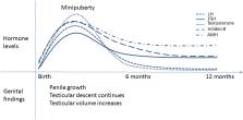
          Cryptorchidism, or undescended testis, is a well-known risk factor for testicular cancer and impaired semen quality in adulthood, conditions which have their origins in early fetal and postnatal life. In human pregnancy, the interplay of testicular and placental hormones as well as local regulatory factors and control by the hypothalamic-pituitary (HP) axis, lead to testicular descent by term. The normal masculine development may be disrupted by environmental factors or genetic defects and result in undescended testes. Minipuberty refers to the postnatal re-activation of the HP-testicular (T) axis after birth. During the first weeks of life, gonadotropin levels increase, followed by activation and proliferation of testicular Leydig, Sertoli and germ cells. Consequent rise in testosterone levels results in penile growth during the first months of life. Testicular size increases and testicular descent continues until three to five months of age. Insufficient HPT axis activation (e.g., hypogonadotropic hypogonadism) is often associated with undescended testis and therefore minipuberty is considered an important phase in the normal male reproductive development. Minipuberty provides a unique window of opportunity for the early evaluation of HPT axis function during early infancy. For cryptorchid boys, hormonal evaluation during minipuberty may give a hint of the underlying etiology and aid in the evaluation of the later risk of HPT axis dysfunction and impaired fertility. The aim of this review is to summarize the current knowledge of the role of minipuberty in testicular development and descent.

          Related collections

          Most cited references114

          • Record: found
          • Abstract: found
          • Article: not found

          Identification in rats of a programming window for reproductive tract masculinization, disruption of which leads to hypospadias and cryptorchidism.

          Becoming a phenotypic male is ultimately determined by androgen-induced masculinization. Disorders of fetal masculinization, resulting in hypospadias or cryptorchidism, are common, but their cause remains unclear. Together with the adult-onset disorders low sperm count and testicular cancer, they can constitute a testicular dysgenesis syndrome (TDS). Although masculinization is well studied, no unifying concept explains normal male reproductive development and its abnormalities, including TDS. We exposed rat fetuses to either anti-androgens or androgens and showed that masculinization of all reproductive tract tissues was programmed by androgen action during a common fetal programming window. This preceded morphological differentiation, when androgen action was, surprisingly, unnecessary. Only within the programming window did blocking androgen action induce hypospadias and cryptorchidism and altered penile length in male rats, all of which correlated with anogenital distance (AGD). Androgen-driven masculinization of females was also confined to the same programming window. This work has identified in rats a common programming window in which androgen action is essential for normal reproductive tract masculinization and has highlighted that measuring AGD in neonatal humans could provide a noninvasive method to predict neonatal and adult reproductive disorders. Based on the timings in rats, we believe the programming window in humans is likely to be 8-14 weeks of gestation.
            Bookmark
            • Record: found
            • Abstract: found
            • Article: not found

            Kisspeptin neurons in the arcuate nucleus of the ewe express both dynorphin A and neurokinin B.

            Kisspeptin is a potent stimulator of GnRH secretion that has been implicated in the feedback actions of ovarian steroids. In ewes, the majority of hypothalamic kisspeptin neurons are found in the arcuate nucleus (ARC), with a smaller population located in the preoptic area. Most arcuate kisspeptin neurons express estrogen receptor-alpha, as do a set of arcuate neurons that contain both dynorphin and neurokinin B (NKB), suggesting that all three neuropeptides are colocalized in the same cells. In this study we tested this hypothesis using dual immunocytochemistry and also determined if kisspeptin neurons contain MSH or agouti-related peptide. To assess colocalization of kisspeptin and dynorphin, we used paraformaldehyde-fixed tissue from estrogen-treated ovariectomized ewes in the breeding season (n = 5). Almost all ARC, but no preoptic area, kisspeptin neurons contained dynorphin. Similarly, almost all ARC dynorphin neurons contained kisspeptin. In experiment 2 we examined colocalization of kisspeptin and NKB in picric-acid fixed tissue collected from ovary intact ewes (n = 9). Over three quarters of ARC kisspeptin neurons also expressed NKB, and a similar percentage of NKB neurons contained kisspeptin. In contrast, no kisspeptin neurons stained for MSH or agouti-related peptide. These data demonstrate that, in the ewe, a high percentage of ARC kisspeptin neurons also produce dynorphin and NKB, and we propose that a single subpopulation of ARC neurons contains all three neuropeptides. Because virtually all of these neurons express estrogen and progesterone re-ceptors, they are likely to relay the feedback effects of these steroids to GnRH neurons to regulate reproductive function.
              Bookmark
              • Record: found
              • Abstract: found
              • Article: not found

              Difference in prevalence of congenital cryptorchidism in infants between two Nordic countries.

              Several investigators have shown striking differences in semen quality and testicular cancer rate between Denmark and Finland. Since maldescent of the testis is a shared risk factor for these conditions we undertook a joint prospective study for the prevalence of congenital cryptorchidism. 1068 Danish (1997-2001) and 1494 Finnish boys (1997-99) were consecutively recruited prenatally. We also established prevalence data for all newborns at Turku University Central Hospital, Finland (1997-99, n=5798). Testicular position was assessed by a standardised technique. All subtypes of congenital cryptorchidism were included, but retractile testes were considered normal. Prevalence of cryptorchidism at birth was 9.0% (95% CI 7.3-10.8) in Denmark and 2.4% (1.7-3.3) in Finland. At 3 months of age, prevalence rates were 1.9% (1.2-3.0) and 1.0% (0.5-1.7), respectively. Significant geographic differences were still present after adjustment for confounding factors (birthweight, gestational age, being small for gestational age, maternal age, parity, mode of delivery); odds ratio (Denmark vs Finland) was 4.4 (2.9-6.7, p<0.0001) at birth and 2.2 (1.0-4.5, p=0.039) at three months. The rate in Denmark was significantly higher than that reported 40 years ago. Our findings of increasing and much higher prevalence of congenital cryptorchidism in Denmark than in Finland contribute evidence to the pattern of high frequency of reproductive problems such as testicular cancer and impaired semen quality in Danish men. Although genetic factors could account for the geographic difference, the increase in reproductive health problems in Denmark is more likely explained by environmental factors, including endocrine disrupters and lifestyle.
                Bookmark

                Author and article information

                Contributors
                Journal
                Front Endocrinol (Lausanne)
                Front Endocrinol (Lausanne)
                Front. Endocrinol.
                Frontiers in Endocrinology
                Frontiers Media S.A.
                1664-2392
                23 July 2019
                2019
                : 10
                : 489
                Affiliations
                [1] 1Department of Pediatrics, Kuopio University Hospital , Kuopio, Finland
                [2] 2Research Centre for Integrative Physiology and Pharmacology, Institute of Biomedicine, University of Turku , Turku, Finland
                [3] 3Department of Pediatrics, Turku University Hospital , Turku, Finland
                [4] 4Barts and the London, William Harvey Research Institute, Queen Mary University of London , London, United Kingdom
                Author notes

                Edited by: Richard Quinton, Newcastle University, United Kingdom

                Reviewed by: Kate Lakoski Loveland, Monash University, Australia; Ellis Fok, The Chinese University of Hong Kong, China

                *Correspondence: Tanja Kuiri-Hänninen tanja.kuiri-hanninen@ 123456kuh.fi

                This article was submitted to Reproduction, a section of the journal Frontiers in Endocrinology

                †These authors share first authorship

                Article
                10.3389/fendo.2019.00489
                6663997
                31396156
                b0f9ad81-6b05-4312-9e0e-4634e51da34f
                Copyright © 2019 Kuiri-Hänninen, Koskenniemi, Dunkel, Toppari and Sankilampi.

                This is an open-access article distributed under the terms of the Creative Commons Attribution License (CC BY). The use, distribution or reproduction in other forums is permitted, provided the original author(s) and the copyright owner(s) are credited and that the original publication in this journal is cited, in accordance with accepted academic practice. No use, distribution or reproduction is permitted which does not comply with these terms.

                History
                : 16 April 2019
                : 04 July 2019
                Page count
                Figures: 2, Tables: 1, Equations: 0, References: 123, Pages: 12, Words: 10002
                Categories
                Endocrinology
                Mini Review

                Endocrinology & Diabetes
                hpg axis,cryptorchidism,minipuberty of infancy,testicular descent,gonadotropin (fsh and lh)

                Comments

                Comment on this article