8
views
0
recommends
+1 Recommend
0 collections
    0
    shares
      • Record: found
      • Abstract: found
      • Article: found
      Is Open Access

      Shenfu Injection attenuates rat myocardial hypertrophy by up-regulating miR-19a-3p expression

      research-article

      Read this article at

      Bookmark
          There is no author summary for this article yet. Authors can add summaries to their articles on ScienceOpen to make them more accessible to a non-specialist audience.

          Abstract

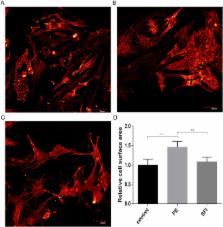
          Shenfu Injection (SFI) is a classical Chinese medicine used to treat heart failure. Our previous study demonstrated that miRNAs underwent changes in rats with myocardial hypertrophy induced by abdominal aortic constriction. Interestingly, there was a significant change in miR-19a-3p, whose target gene is known to be associated with MEF2 signaling. However, whether and how SFI regulates miR-19a-3p in the treatment of myocardial hypertrophy has not been investigated. The purpose of the present study was to investigate the regulatory effect of SFI on miR-19a-3p in MEF2 signaling in the rat hypertrophic myocardium. We found that the miR-19a-3p expression level was significantly decreased in the hypertrophic myocardium, and MEF2A was the target gene of miR-19a-3p. The protein expressions of MEF2A, β-MHC, BNP and TRPC1 were significantly increased in hypertrophic cardiomyocytes. MiR-19a-3p was up-regulated after SFI treatment, and the protein expressions of these genes were significantly decreased. In addition, miR-19a-3p over-expression in hypertrophic cardiomyocytes could decrease MEF2A mRNA and protein expressions, and anti-miR-19a-3p showed the opposite result. Our study provided substantial evidence that miR-19a-3p played a functional role in MEF2 signaling in myocardial hypertrophy. SFI attenuated cardiomyocyte hypertrophy probably through up-regulating or maintaining the miR-19a-3p levels and regulating the MEF2 signaling pathway.

          Related collections

          Most cited references24

          • Record: found
          • Abstract: found
          • Article: not found

          MicroRNA-208a is a regulator of cardiac hypertrophy and conduction in mice.

          MicroRNAs (miRNAs) are a class of small noncoding RNAs that have gained status as important regulators of gene expression. Here, we investigated the function and molecular mechanisms of the miR-208 family of miRNAs in adult mouse heart physiology. We found that miR-208a, which is encoded within an intron of alpha-cardiac muscle myosin heavy chain gene (Myh6), was actually a member of a miRNA family that also included miR-208b, which was determined to be encoded within an intron of beta-cardiac muscle myosin heavy chain gene (Myh7). These miRNAs were differentially expressed in the mouse heart, paralleling the expression of their host genes. Transgenic overexpression of miR-208a in the heart was sufficient to induce hypertrophic growth in mice, which resulted in pronounced repression of the miR-208 regulatory targets thyroid hormone-associated protein 1 and myostatin, 2 negative regulators of muscle growth and hypertrophy. Studies of the miR-208a Tg mice indicated that miR-208a expression was sufficient to induce arrhythmias. Furthermore, analysis of mice lacking miR-208a indicated that miR-208a was required for proper cardiac conduction and expression of the cardiac transcription factors homeodomain-only protein and GATA4 and the gap junction protein connexin 40. Together, our studies uncover what we believe are novel miRNA-dependent mechanisms that modulate cardiac hypertrophy and electrical conduction.
            Bookmark
            • Record: found
            • Abstract: found
            • Article: not found

            Molecular distinction between physiological and pathological cardiac hypertrophy: experimental findings and therapeutic strategies.

            Cardiac hypertrophy can be defined as an increase in heart mass. Pathological cardiac hypertrophy (heart growth that occurs in settings of disease, e.g. hypertension) is a key risk factor for heart failure. Pathological hypertrophy is associated with increased interstitial fibrosis, cell death and cardiac dysfunction. In contrast, physiological cardiac hypertrophy (heart growth that occurs in response to chronic exercise training, i.e. the 'athlete's heart') is reversible and is characterized by normal cardiac morphology (i.e. no fibrosis or apoptosis) and normal or enhanced cardiac function. Given that there are clear functional, structural, metabolic and molecular differences between pathological and physiological hypertrophy, a key question in cardiovascular medicine is whether mechanisms responsible for enhancing function of the athlete's heart can be exploited to benefit patients with pathological hypertrophy and heart failure. This review summarizes key experimental findings that have contributed to our understanding of pathological and physiological heart growth. In particular, we focus on signaling pathways that play a causal role in the development of pathological and physiological hypertrophy. We discuss molecular mechanisms associated with features of cardiac hypertrophy, including protein synthesis, sarcomeric organization, fibrosis, cell death and energy metabolism and provide a summary of profiling studies that have examined genes, microRNAs and proteins that are differentially expressed in models of pathological and physiological hypertrophy. How gender and sex hormones affect cardiac hypertrophy is also discussed. Finally, we explore how knowledge of molecular mechanisms underlying pathological and physiological hypertrophy may influence therapeutic strategies for the treatment of cardiovascular disease and heart failure. 2010 Elsevier Inc. All rights reserved.
              Bookmark
              • Record: found
              • Abstract: found
              • Article: not found

              Hypertrophy of the heart: a new therapeutic target?

              Recent studies call into question the necessity of hypertrophic growth of the heart as a "compensatory" response to hemodynamic stress. These findings, coupled with recent progress in dissecting the molecular bases of hypertrophy, raise the prospect of suppressing hypertrophy without provoking circulatory insufficiency. In this article, we focus on signaling pathways that hold promise as potential targets for therapeutic intervention. We also summarize observations from animal models and clinical trials that suggest benefit from an antihypertrophic strategy.
                Bookmark

                Author and article information

                Contributors
                yuanwj@hotmail.com
                qinsmmu@126.com
                Journal
                Sci Rep
                Sci Rep
                Scientific Reports
                Nature Publishing Group UK (London )
                2045-2322
                16 March 2018
                16 March 2018
                2018
                : 8
                : 4660
                Affiliations
                [1 ]ISNI 0000 0000 8744 8924, GRID grid.268505.c, Department of Pharmacognosy, , Zhejiang Chinese Medical University School of Pharmacy, ; Hangzhou, 310053 ZJ China
                [2 ]ISNI 0000 0004 1761 9803, GRID grid.412194.b, Department of Physiology, , Ningxia Medical University, ; Yinchuan, 750004 NX China
                [3 ]ISNI 0000 0004 0369 1660, GRID grid.73113.37, Department of Physiology, , Second Military Medical University, ; Shanghai, 200433 China
                Article
                23137
                10.1038/s41598-018-23137-4
                5856750
                29549288
                b26e47de-0cc2-4e3a-8226-31df08ce0cde
                © The Author(s) 2018

                Open Access This article is licensed under a Creative Commons Attribution 4.0 International License, which permits use, sharing, adaptation, distribution and reproduction in any medium or format, as long as you give appropriate credit to the original author(s) and the source, provide a link to the Creative Commons license, and indicate if changes were made. The images or other third party material in this article are included in the article’s Creative Commons license, unless indicated otherwise in a credit line to the material. If material is not included in the article’s Creative Commons license and your intended use is not permitted by statutory regulation or exceeds the permitted use, you will need to obtain permission directly from the copyright holder. To view a copy of this license, visit http://creativecommons.org/licenses/by/4.0/.

                History
                : 7 August 2017
                : 12 November 2017
                Categories
                Article
                Custom metadata
                © The Author(s) 2018

                Uncategorized
                Uncategorized

                Comments

                Comment on this article