25
views
0
recommends
+1 Recommend
0 collections
    12
    shares
      • Record: found
      • Abstract: found
      • Article: found
      Is Open Access

      Transient Cannabinoid Receptor 2 Blockade during Immunization Heightens Intensity and Breadth of Antigen-specific Antibody Responses in Young and Aged mice

      research-article

      Read this article at

      Bookmark
          There is no author summary for this article yet. Authors can add summaries to their articles on ScienceOpen to make them more accessible to a non-specialist audience.

          Abstract

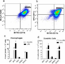
          The hallmark of vaccines is their ability to prevent the spread of infectious pathogens and thereby serve as invaluable public health tool. Despite their medical relevance, there is a gap in our understanding of the physiological factors that mediate innate and adaptive immune response to vaccines. The endocannabinoid (eCB) system is a critical modulator of homeostasis in vertebrates. Our results indicate that macrophages and dendritic cells produce the endocannabinoid, 2-arachidonoyl-sn-glycerol (2-AG) upon antigen activation. We have also established that 2-AG levels are upregulated in the serum and in the lymph node of mice during vaccination. We hypothesized that the intrinsic release of eCBs from immune cells during activation by pathogenic antigens mitigate inflammation, but also suppress overall innate and adaptive immune response. Here we demonstrate, for the first time, that transient administration of the cannabinoid receptor 2 antagonist AM630 (10 mg/kg) or inverse agonist JTE907 (3 mg/kg) during immunization heightens the intensity and breadth of antigen-specific immune responses in young and aged mice through the upregulation of immunomodulatory genes in secondary lymphoid tissues.

          Related collections

          Most cited references61

          • Record: found
          • Abstract: found
          • Article: not found

          Expression of central and peripheral cannabinoid receptors in human immune tissues and leukocyte subpopulations.

          Two proteins with seven transmembrane-spanning domains typical of guanosine-nucleotide-binding-protein-coupled receptors have been identified as cannabinoid receptors; the central cannabinoid receptor, CB1, and the peripheral cannabinoid receptor, CB2, initially described in rat brain and spleen, respectively. Here, we report the distribution patterns for both CB1 and CB2 transcripts in human immune cells and in several human tissues, as analysed using a highly sensitive and quantitative PCR-based method. CB1 was mainly expressed in the central nervous system and, to a lower extent, in several peripheral tissues such as adrenal gland, heart, lung, prostate, uterus, ovary, testis, bone marrow, thymus and tonsils. In contrast, the CB2 gene, which is not expressed in the brain, was particularly abundant in immune tissues, with an expression level 10-100-fold higher than that of CB1. Although CB2 mRNA was also detected in some other peripheral tissues, its level remained very low. In spleen and tonsils, the CB2 mRNA content was equivalent to that of CB1 mRNA in the central nervous system. Among the main human blood cell subpopulations, the distribution pattern of the CB2 mRNA displayed important variations. The rank order of CB2 mRNA levels in these cells was B-cells > natural killer cells > monocytes > polymorphonuclear neutrophil cells > T8 cells > T4 cells. The same rank order was also established in human cell lines belonging to the myeloid, monocytic and lymphoid lineages. The prevailing expression of the CB2 gene in immune tissues was confirmed by Northern-blot analysis. In addition, the expression of the CB2 protein was demonstrated by an immunohistological analysis performed on tonsil sections using specific anti-(human CB2) IgG; this experiment showed that CB2 expression was restricted to B-lymphocyte-enriched areas of the mantle of secondary lymphoid follicles. These results suggest that (a) CB1 and CB2 can be considered as tissue-selective antigens of the central nervous system and immune system, respectively, and (b) cannabinoids may exert specific receptor-mediated actions on the immune system through the CB2 receptor.
            Bookmark
            • Record: found
            • Abstract: found
            • Article: not found

            Granulocyte-macrophage colony-stimulating factor (CSF) and macrophage CSF-dependent macrophage phenotypes display differences in cytokine profiles and transcription factor activities: implications for CSF blockade in inflammation.

            GM-CSF and M-CSF (CSF-1) can enhance macrophage lineage numbers as well as modulate their differentiation and function. Of recent potential significance for the therapy of inflammatory/autoimmune diseases, their blockade in relevant animal models leads to a reduction in disease activity. What the critical actions are of these CSFs on macrophages during inflammatory reactions are unknown. To address this issue, adherent macrophages (GM-BMM and BMM) were first derived from murine bone marrow precursors by GM-CSF and M-CSF, respectively, and stimulated in vitro with LPS to measure secreted cytokine production, as well as NF-kappaB and AP-1 activities. GM-BMM preferentially produced TNF-alpha, IL-6, IL-12p70, and IL-23 whereas, conversely, BMM generated more IL-10 and CCL2; strikingly the latter population could not produce detectable IL-12p70 and IL-23. Following LPS stimulation, GM-BMM displayed rapid IkappaBalpha degradation, RelA nuclear translocation, and NF-kappaB DNA binding relative to BMM, as well as a faster and enhanced AP-1 activation. Each macrophage population was also pretreated with the other CSF before LPS stimulation and found to adopt the phenotype of the other population to some extent as judged by cytokine production and NF-kappaB activity. Thus, GM-CSF and M-CSF demonstrate, at the level of macrophage cytokine production, different and even competing responses with implications for their respective roles in inflammation, including a possible dampening or suppressive role for M-CSF in certain circumstances.
              Bookmark
              • Record: found
              • Abstract: found
              • Article: not found

              Chemokine Requirements for B Cell Entry to Lymph Nodes and Peyer's Patches

              B cell entry to lymph nodes and Peyer's patches depends on chemokine receptor signaling, but the principal chemokine involved has not been defined. Here we show that the homing of CXCR4−/− B cells is suppressed in CCL19 (ELC)- and CCL21 (SLC)-deficient paucity of lymph node T cells mice, but not in wild-type mice. We also find that CXCR4 can contribute to T cell homing. Using intravital microscopy, we find that B cell adhesion to high endothelial venules (HEVs) is disrupted when CCR7 and CXCR4 are predesensitized. In Peyer's patches, B cell entry is dependent on CXCR5 in addition to CCR7/CXCR4. CXCL12 (SDF1) is displayed broadly on HEVs, whereas CXCL13 (BLC) is found selectively on Peyer's patch follicular HEVs. These findings establish the principal chemokine and chemokine receptor requirements for B cell entry to lymph nodes and Peyer's patches.
                Bookmark

                Author and article information

                Journal
                Sci Rep
                Sci Rep
                Scientific Reports
                Nature Publishing Group
                2045-2322
                17 February 2017
                2017
                : 7
                : 42584
                Affiliations
                [1 ]Division of Infectious Diseases, Department of Medicine, University of California Irvine , Irvine, CA, USA
                [2 ]Department of Pharmaceutical Sciences, University of California Irvine , Irvine, CA, USA
                [3 ]Division of Biomedical Sciences, School of Medicine, University of California Riverside , Riverside, CA, USA
                [4 ]Department of Physiology & Biophysics, University of California Irvine , Irvine CA, USA
                [5 ]Malaria Infection Biology and Immunity Unit, Laboratory of Immunogenetics, NIAID, NIH , Rockville, MD, USA.
                Author notes
                Article
                srep42584
                10.1038/srep42584
                5314369
                28209996
                b2752345-7396-4468-97c1-cea980767072
                Copyright © 2017, The Author(s)

                This work is licensed under a Creative Commons Attribution 4.0 International License. The images or other third party material in this article are included in the article’s Creative Commons license, unless indicated otherwise in the credit line; if the material is not included under the Creative Commons license, users will need to obtain permission from the license holder to reproduce the material. To view a copy of this license, visit http://creativecommons.org/licenses/by/4.0/

                History
                : 31 August 2016
                : 12 January 2017
                Categories
                Article

                Uncategorized
                Uncategorized

                Comments

                Comment on this article