- Record: found
- Abstract: found
- Article: found
Turkish Society of Cardiology consensus report on the rational use of cardiac troponins in daily practice
other
Kaan Okyay
1 ,
Beste Özben Sadıç
2 ,
Asife Şahinarslan
3 ,
Murtaza Emre Durakoğlugil
4 ,
Can Yücel Karabay
5 ,
Semiha Emel Eryüksel
6 ,
Özlem Gülbahar
7 ,
Abdullah Tekin
8 ,
Aylin Yıldırır
1 ,
Bülent Görenek
9 ,
Oğuz Yavuzgil
10 ,
Ali Serdar Fak
2
19 April 2019
Read this article at
There is no author summary for this article yet. Authors can add summaries to their articles on ScienceOpen to make them more accessible to a non-specialist audience.
Abstract
Background and aim of the document
Cardiac troponins (cTn T and I) are protein molecules that are part of the contractile
apparatus of the cardiac muscle. Increase in these biomarkers represents injury of
myocardial cells without giving any evidence for the underlying mechanism (1, 2).
In the latest (fourth) Expert Consensus Document of Universal Definition of Myocardial
Infarction (2), it has been emphasized that the clinical definition of myocardial
infarction (MI, types 1, 2, and 3) refers to the presence of acute myocardial injury
detected by typical rise and/or fall of cTn in the setting of acute myocardial ischemia
evidenced by at least one of the following findings:
Ischemic symptoms
New alterations in electrocardiography (ECG) related with ischemia
Imaging evidence of new loss of viable myocardium or new regional wall motion abnormality
Demonstration of coronary thrombus
Cardiac troponins are also the mainstay for diagnostic algorithms of acute chest pain
as explained in the latest European Society of Cardiology (ESC) Guideline to manage
acute coronary syndromes (ACS) in patients presenting without persistent ST-segment
elevation (3).
Troponins have typically been used for the diagnosis and prognosis of ACS, but they
may be elevated in many stable and unstable cardiac and non-cardiac conditions. The
clinicians, mainly the cardiologists, emergency and intensive care physicians, and
family physicians, should be aware of all these entities. The use of new-generation
high-sensitivity cTn assays have lowered the diagnostic threshold (specificity) leading
to overdiagnoses of patients with ACS, a vast number of cardiac consultations and
inappropriate coronary angiograms or unnecessary hospitalizations causing increased
complications and cost. The underlying mechanisms and clinical significance of troponin
elevations in some cardiac and non-cardiac conditions have not been completely elucidated.
Additionally, many clinicians are not aware of the biochemical assay problems and
pre-analytical and analytical factors that may result false-positive troponin measurements
(4, 5).
Consequently, there is a need for an up-to-date consensus paper systematically explaining
the causes of cTn elevations to make an accurate differential diagnosis. This document
does not aim to evaluate cTn only in acute chest pain. It will first address the identification
of pre-analytical and analytical factors affecting cTn measurement; then it will discuss
cardiac and non-cardiac causes of acute and chronic cTn elevations. The potential
underlying mechanisms and clinical and prognostic significance of troponin elevations
will be emphasized. We sought to represent messages and recommendations for daily
practice with a multidisciplinary approach.
Definition of myocyte injury using cardiac troponins
The diagnosis of MI requires assessment of ischemic symptoms, ECG changes, patient
characteristics, and biochemical evidence of myocardial injury. Any molecule should
be highly specific and sensitive for myocardial injury to be used as a cardiac biomarker.
Since their identification, cTn has become the mainstay for definition of myocardial
injury. While both skeletal and cardiac myocytes possess troponin, troponin I (TnI)
and troponin T (TnT) have distinct cardiac and skeletal isoforms, whereas troponin
C (TnC) is shared in both tissues (6). Therefore, assays for cTnI and cTnT that target
the most stable regions were developed (7). With the advent of high-sensitive cTn
assays, there has been a shift in evaluation of troponin test from binary (negative-positive)
results to highly quantitative assays (8). However, improved sensitivity has identified
several non-ischemic cardiac and non-cardiac conditions that have cTn concentrations
above the 99th percentile, although not as high as expected with a major coronary
occlusion (9). The definition of MI has been modified over the years because of these
advances, and the fourth Universal Definition of MI has been recently published (2).
Detection of an elevated cTn value above the 99th percentile upper reference limit
is acknowledged as myocardial injury. The injury is considered acute if there is a
rise and/or fall of cTn values, and chronic in the event of persistent cTn elevations.
Kinetics of cardiac troponins
In patients with an acute MI, myocyte necrosis ensues after 15 min of ischemia, and
troponins become elevated 2–4 h after symptom onset and peak at 24–48 h (10). Early
observations revealed an initial peak of troponins followed by sustained elevation
lasting up to 14 days. This initial peak was evident only in patients with successful
reperfusion suggesting a biphasic release (11). The explanation for biphasic profile
depends on subcellular localization of cTn. Cardiac troponins are attached to cardiac
myofibrils via tropomyosin. Approximately 6%–8% of cTnT and 2%–4% of cTnI is loosely
bound that form the cytosolic pool (or the early-release pool), and the remaining
cTn is bound as a ternary complex (12). Following myocardial necrosis, cytosolic pool
including free Tn is released first that forms the initial peak; continuous degradation
of structurally bound Tn (structural pool) contributes to sustained release of binary
and ternary complexes, which is also related to infarct size. Since the cytosolic
pool of cTnI is smaller, biphasic profile of cTnI is not as prominent as cTnT (13).
In addition, both binary and ternary forms may undergo post-translational modifications
in the cytosol, and are susceptible to proteolysis and oxidation in the circulation.
Current assays detect all these different forms, so these changes do not affect sensitivity
(7).
Pre-analytical and analytical factors affecting troponin measurement
Alterations of the assays
Because of their myocardial tissue specificity, cTnI and cTnT are accepted as gold
standard biomarkers to detect myocardial injury (2). For cTn measurement, numerous
contemporary and bedside [point of care (POC)] tests have been approved. In clinical
practice, standardizing of cTnI assays is not feasible leading to non-identical values,
since different antibodies recognize different epitopes of cTnI. In contrast to cTnI,
in cTnT assays, same antibodies are used. However, because of use of different calibrations,
the reported values are not the same between fourth and fifth generation (high-sensitive,
hs) assays (14, 15). To overcome the equivocacy of assay-to-assay differences, direct
comparisons of approved cTn assays are needed (14).
There are some terms used in determination of the analytical view of cTn assays:
Coefficient of variation (CV) is an estimation of reproducibility of the test (day-to-day
imprecision), and it is calculated as the ratio of the standard deviation over the
mean value for repeated testing of the same sample over multiple days (14, 16).
Limit of detection (LOD) is the lowest value measured by progressively dilutions,
and used for ruling out MI (14, 17).
The upper reference limit (URL) is the upper limit of the population of interest,
and is defined as the 99th percentile of the normal distribution. Hence, 1% of otherwise
healthy subjects may still have a cTn value higher than the 99th percentile URL (16).
High-sensitivity assays should have a CV of <10% at the 99th percentile value. Additionally,
with hs assays, concentrations above the LOD, but below the 99th percentile should
be detectable for at least >50% of healthy individuals (2, 14).
The use of non-hs-cTn assays without <10% CV at the 99th percentile URL makes the
monitoring of significant changes more difficult, but it does not result in false-positive
results. Assays with CVs between 10% and 20% are acceptable for clinical use. However,
assays with CVs >20% should not be used. If a cTn assay is not available, the best
option is measurement of CK-MB mass activity. As with hs-cTn assays, an increased
CK-MB denotes a value above the 99th percentile URL (sex-specific URLs are recommended
for both) (2).
Immunoassay interferences
Interferences in immunoassay may cause in confounding results, and they lead the physician
to give inappropriate treatments even to perform unnecessary interventions. Interfering
substances may result in falsely elevated or falsely low measurements in different
assay systems. Laboratories should detect, test, and report suspected interferences.
It is of great importance to communicate with the laboratory for any discordance between
the clinical characteristics and the laboratory data (18).
Pre-analytical interferences
Serum, plasma, and anticoagulated whole blood are available specimens for the analyses
(14). When hs-cTnI assays are utilized, important differences can be obtained using
different samples (serum vs. heparin plasma vs. EDTA plasma) (19).
Several cTn assays are influenced by hemolysis dependent of the amount of cTn and
free hemoglobin concentrations. Some assays reported decreased results, and the others
are either unaffected by hemolysis or reported falsely elevated results (19). In this
point, clinicians should view the properties of the commercial kits in coordination
with central laboratory.
Fibrin may be present in the blood collection tubes as visible clot or invisible strands.
These fibrin substances may affect cTn assays by interfering with antigen-antibody
binding. Fibrin strands can be eliminated if the recommended times subsequent centrifugations
are appointed (18).
Antibody interferences
Both heterophile and anti-animal antibodies may result in immunoassay interferences.
Heterophile antibodies are emerged against many antigens that are not clearly determined.
The differentiation of these antibodies is not always possible (20). Autoimmune antibodies
can cause immunoassay interferences. In many cases, a specific antigen is not well
defined. However, in some cases, antibody production occurs because of exposure to
intestinal and pulmonary bacteria. Even dietary proteins, vaccines, and multiparity
may be responsible for this reaction. Interfering antibodies are detected more in
males, and they have been shown to rise in response to blood transfusions and exposure
to foreign proteins. The presence of RF in blood samples of the patients either with
rheumatic or with non-rheumatic diseases is responsible for false-positive troponin
assays (20). Human anti-animal antibodies are specific polyclonal antibodies against
to specific animal immunogens, most commonly to mouse, but also rat, rabbit, goat,
sheep, pig, cattle, and horse antigens (18, 20). These antibodies are also found in
subjects having contact with domestic animals such as cat and dog.
Initially, the prevalence of interfering antibodies was reported between 10% and 40%
depending on the assay and population of interest. However, the newer assays containing
blocking agents added to reagents have lowered the ratio of interference to less than
2% (20). In addition, increased activity of endogenous alkaline phosphatase (ALP),
or treatment with exogenous ALP (asfotase) in patients with phosphatasia may cause
interference with some cTnI assays (21, 22).
There are some techniques to deal with antibody interferences. A simple method is
the analysis of the sample using an alternate assay. Another procedure is measurement
before and after applying of a blocking reagent or using heterophile-blocking tubes.
Repeated measurement with manufacturer’s diluents is another option. An anti-animal
interference can also be eliminated by precipitation with polyethylene glycol precipitation
(PEG 6000) (18).
The conditions related with elevation in cardiac troponins
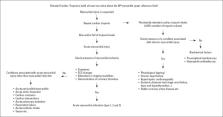
There is a wide range of conditions related with elevated cTn. In Figure 1, we have
summarized reasons for acute and chronic elevations. From our point of view, classifying
of these conditions as cardiac and non-cardiac and as stable and unstable can be instructive
for understanding the underlying mechanisms and helpful in decision making. In this
sense, first, we explain the stable and unstable cardiac and non-cardiac conditions.
Later, we propose an algorithm for management of elevated cTn (Fig. 2). We strongly
recommend against using the term of “troponinemia”. Any increase in the troponins
needs to be taken into account and should be monitored. When persistently elevated
cTn levels (that means ≤20% variation) are detected, conditions related with chronic
cardiac injury should be regarded. When these conditions are excluded, biochemical
factors could be the culprit. A typical rise and/or fall of troponin levels (with
at least one value above the 99th percentile upper reference limit) after repeated
measure (preferably 3 h later) is called as acute myocardial injury, which is further
defined as acute MI when accompanied by myocardial ischemia; otherwise, unstable cardiac
and non-cardiac conditions should be considered.
Figure 1
Schematic presentation of the conditions related with elevated cardiac troponins
Figure 2
Algorithm for the management of the cardiac troponin elevation
Stable cardiac conditions
Chronic heart failure
In patients with advanced heart failure (HF), elevated troponin levels are commonly
observed and are indicative of adverse prognosis (23). Increased volume and pressure
load results in myocardial wall strain and myocyte death that are accepted to be the
underlying mechanisms (23, 24). The relationship between wall strain and myocyte death
could be explained by impaired subendocardial perfusion leading to cell death (25).
Increased brain natriuretic peptide (BNP) level, which is an indicator for myocardial
strain, correlates with increased troponin levels (26). Moreover, in rat myocardium,
increased strain resulted in troponin elevation regardless of ischemia (27). Myocardial
cell loss is considered the main underlying mechanism in progression of advanced HF.
Sympathetic system and RAS activation, inflammatory mediators, and increased integrin
levels as well as oxidative stress may enhance myocardial loss in patients with HF
(28, 29). Increased troponin levels were also associated with acute decompensation,
progressive disease, and poor prognosis in acute and chronic HF (30, 31).
Hypertrophic cardiomyopathy
In hypertrophic cardiomyopathy (HCMP), troponin rise may be seen because of several
factors such as increased myocardial volume, increased oxygen need, and decreased
flow volume because of remodeling (32). Increased serum troponin level observed in
a significant ratio of the patients with HCMP and is an independent predictor of adverse
outcome (33). Elevated troponin levels have also been detected in other structural
heart diseases associated with left ventricular wall thickening.
Infiltrative cardiac disorders
According to the accumulating substance, some of the infiltrative cardiac diseases
increase ventricular wall thickness, while others cause chamber enlargement with secondary
wall thinning. Cardiac amyloidosis is a primary restrictive cardiomyopathy. Although
pathophysiology of troponin elevation remains unclear, myocyte compression injury
due to extracellular deposition of the amyloid plaque is held to be responsible (34).
Dispenzieri et al. (35) showed that in cardiac amyloidosis, troponin levels are more
valuable as survival predictors than ECG and symptoms. Likewise, hs-TnT value in sarcoidosis
is considered an important marker of disease activity and is decreased following steroid
therapy (36).
Consensus Statements for Elevation of Cardiac Troponins
References
Cardiac troponins (cTn) are the mainstay for definition of myocardial injury.
1-3
Cardiac troponins should be used for the diagnosis and prognosis of acute coronary
syndromes.
2, 3
An elevated troponin level should always be interpreted in the context of the clinical
presentation
• Typical rise and fall, with at least one value above the 99th percentile URL, in
the setting of acute myocardial ischemia (evidenced by ischemic symptoms, and alterations
in ECG or imaging modalities) are needed for an accurate diagnosis of myocardial infarction.
• Troponin follow-up along with symptoms and ECG changes are required in case of suspicion
2, 3
A “sole” troponin elevation should not be defined as myocardial infarction.
2, 3
Troponin elevation is not recommended for the purpose of identification of the etiology
of myocardial injury.
2, 3
Stable coronary artery disease, chronic heart failure, acute pericarditis, myocarditis,
Takatsubo syndrome, tachycardia, cardiac interventions should be considered among
the stable and non-stable cardiac conditions of increased troponins (generally with
different enzyme kinetics).
2, 30, 44, 45, 51, 61, 73, 82, 88
Aging, renal failure, sepsis, pulmonary hypertension, acute pulmonary embolism, critically
ill patients, acute cerebrovascular events should be considered among the stable and
non-stable non-cardiac conditions of increased troponins (generally with different
enzyme kinetics).
92, 96, 100, 113, 117, 123
Troponin elevation is prognostic even when ACS is excluded.
• Troponin levels are recommended to predict prognosis in patients with heart failure,
stable chest pain, chronic renal disease, pulmonary diseases, stroke or after cardiac
interventions and cardiac surgery. • Troponins may be used as surrogate markers of
coronary artery disease mortality for screening and monitoring of healthy subjects.
30, 49-51, 96, 111, 116, 125
Numerous assays are approved for troponin measurement.
• 99th percentile for high-sensitive (hs) cTn assays should be measured with an analytical
imprecision (coefficient of variation -CV-) of 10% or less. • Non-hs-cTn assays with
CV between 10% and 20% may also be used. • Assays with CVs >20% should not be used.
• Measurement of CK-MB mass activity may be used if a cTn assay is not available.
• Analysis of the sample using an alternate cTn assay, measurement before and after
using of heterophile-blocking tubes, and measurement of a series of dilutions may
be used to deal with laboratory interferences related with false- positive results.
• Physicians should communicate with the laboratory when a persistent cTn elevation
(that means ≤20% variation by repeated measures) cannot be explained by any clinical
condition.
14-16, 18-20
Turnover of myocardial cells, apoptosis
Troponin values increase generally because of myocyte necrosis caused by ischemia
and MI. In some cases, an increase in troponin levels may be seen without myocyte
necrosis. All cells, including cardiomyocytes, have death protocols that are activated
when appropriate conditions are met. These protocols can be activated because of temporary
conditions such as apoptosis, preload increase, ischemia, or pulmonary hypertension
(37). Our present knowledge is not sufficient to determine by which potential effects
apoptosis increases troponin levels (38).
Drug toxicity
Many agents can have cardiotoxic effects. Cardiotoxicity is often observed with use
of anthracyclines, which are effective drugs for the treatment of solid and hematologic
malignancies, and trastuzumab-like drugs, which is an HER-2/neu (Human Epidermal Growth
Factor Receptor 2) receptor antagonist. While cardiotoxic effects of anthracycline
derivatives are dose-dependent and irreversible, trastuzumab-like drugs have reversible
effects. Cell membrane damage, caused by oxidative stress, reactive oxygen species,
and lipid peroxidation, is held responsible for anthracycline group cardiotoxicity.
Anti-HER2 drug group toxicity has reversible, functional, and structural effects on
contractive proteins, and mitochondria, therefore rarely causes cell death (39, 40).
Chemotherapy-induced troponin rise can predict the forthcoming left ventricular (LV)
dysfunction (41, 42).
Cardiac surgery
As in all patient groups, post-operative troponin elevation is associated with poor
prognosis. Troponin level increases >10 times of the 99th percentile URL in patients
with normal baseline values on the first 48 h after “on pump” valve or coronary by-pass
surgery is an important predictor for the first year survival rate (2, 43).
Percutaneous coronary/valvular interventions
Troponin level increase can be observed in percutaneous coronary interventions in
stable settings due to flow discontinuation during balloon dilatation or ischemia
due to distal embolization, and it is indicative of myocyte necrosis. At least five-fold
increase in troponins predicts cardiovascular events at 30 days and one-year follow-up
(2). Troponin level increase can also be observed after percutaneous valvular interventions
such as transcutaneous aortic valve implantation (TAVI). Pre-interventional increased
troponin values are observed in most of the patients undergoing TAVI due to critical
aortic stenosis. Pre- and post-interventional levels are considered important prognostic
factors of one-year survival rate independently from the success of intervention (44).
Cardiac pacing, cardioversion, and ablation therapies
Troponin increase can occur following permanent pacemaker insertion due to minimal
myocardial damage caused during endocardial lead implantation (45). A mild but still
significant increase in troponin levels has been observed following electrical cardioversion
(CV) in non-valvular atrial fibrillation (AF). This increase is even more significant
in patients with increased LV volume and low ejection fraction (EF) (46). Even though
troponin increase may show progression of the present HF status, we must also keep
in mind that this increase can be due to implantable cardioverter defibrillator shocks
in patients with HF (47).
Radiofrequency ablation, which is performed in various arrhythmias such as supraventricular
tachycardia (SVT), AF, and ventricular tachycardia, causes cardiac damage via thermal
energy, therefore causing troponin increase (48).
Stable coronary artery disease
Several clinical studies have demonstrated that troponin value in otherwise healthy
subjects could be a predictor of subsequent adverse cardiac events including mortality
(49, 50). The PROMISE (Prospective Multicenter Imaging Study for Evaluation of Chest
Pain) study has provided that in patients with stable chest pain and suspected coronary
artery disease (CAD), the upper hsTnI quartiles were independently related with death,
acute MI, or hospitalization for unstable chest pain during one-year follow-up period
(51). Despite promising results, further data are required using troponins as surrogate
markers of CAD mortality to screen and monitor the healthy subjects.
Unstable cardiac conditions
Acute coronary syndrome
ACS is a term that includes patients with ST-segment elevation myocardial infarction
(STEMI), non-ST-segment elevation myocardial infarction (NSTEMI), and unstable angina
(UA). Cardiac biomarker elevations are required to distinguish NSTEMI from UA and
helpful in patients with chest pain. Many diagnostic algorithms incorporated with
serial cTn measurement are proposed to rule-in/out in patients with acute chest pain
(3, 52, 53). It should be remembered that some diseases like myocarditis, takotsubo
cardiomyopathy might produce dynamic changes in cTn levels (53); and late presentation
of ACS might not show meaningful changes in cTn.
Rapid rule-in and rule-out strategies for patients admitted with chest pain to the
emergency department use different time points and cut-off values. The latest ESC
Guideline for the management of ACS in patients presenting without persistent ST-segment
elevation (3) depicted a comprehensive algorithm. Briefly, in acute chest pain, either
1-h or 3-h strategy should be used. The 1-h strategy is applicable if chest pain onset
is >3 h, and there is a high pre-test probability for NSTEMI. Additionally, it can
be applied when only high-sensitivity cTn assays are available [hs-cTnT (Elecsys),
hs-cTnI (Architect), and hs-cTnI (Dimension Vista) are the validated hs-cTn assays];
and the cut-off levels are assay specific. The guideline recommended using the 3-h
strategy, in which one delta value is greater than URL during follow-up prompts invasive
strategy. We believe that the 3-h strategy is more user friendly and has a high validity,
and it should be preferred. These strategies have a negative predictive value (for
rule-out) exceeding 98%. On the other hand, the positive predictive value (for rule-in)
is between 75% and 80% (3).
Apart from its diagnostic usefulness, cTn elevation conveys prognostic value. The
patients with elevated cTn have increased mortality rate; and they are likely to have
coronary thrombosis, more complex coronary lesions, and diminished ventricular function
(54, 55).
Severe hypertension
Increased troponin levels can be seen in hypertensive crisis because of supply-demand
mismatch or obstructive CAD. In the study reported by Pattanshetty et al. (56), increased
troponin levels in patients referring to the hospital because of hypertensive crisis
were related with increased adverse cardiac events ratio. Interestingly, in one-fourth
of these patients, obstructive CAD was not seen.
Aortic dissection
Almost 90% of the patients with aortic dissection (AD) have abnormal ECG with 25%–35%
having ACS-like ECG features resembling NSTEMI and 4%–16% have ECG findings of STEMI
(57-60). Troponin positivity was reported from 16% to 33% with standard assays and
54% to 61% with hs assay with no difference between type A and type B dissection.
Troponin elevation in the setting of AD may be due to intimal flap obstructing the
coronary ostia, coronary ostia dissection, decreased blood pressure, aortic regurgitation,
LV pressure and volume overload, increased sympathetic drive leading to microvascular
dysfunction, and preexisting CAD (57-60).
Takotsubo syndrome
Takotsubo syndrome (TTS) is characterized by temporary LV wall motion abnormality
that is usually preceded by emotional or physical triggers. The clinical presentation
of patients with TTS is very similar to those of ACS subjects. The International Takotsubo
Registry (61) found that troponin levels were elevated in 87% of 1750 patients on
admission. Recorded troponin values are disproportionally low considering the extensive
LV involvement (62). The mean troponin levels at admission were found to be similar
to those in patients with ACS. Yet, peak values of cTn are lower in TTS than patients
with ACS, though comparable values with NSTEMI could be observed (61). Troponin peaks
occur earlier, usually at presentation or within 24 h following the onset of symptoms
and normalizes faster than STEMI (63). Higher TnI values were observed in patients
with TTS presenting with cardiogenic shock (64).
Cardiac contusion
Cardiac contusion or currently preferred term blunt cardiac injury (BCI) is usually
suspected in blunt thoracic trauma (BTT). There are no definitive diagnostic criteria,
but various combinations of clinical picture, ECG, troponin, or cardiac imaging were
used to define BCI. Therefore, the incidence of BCI in patients with BTT varies from
3% to 56% (65-67). The release of troponin reflecting myocardial cell injury is believed
to be because of mechanical transmission of force through the chest wall (68). But,
troponin elevation was also observed in 25%–35% of trauma subjects who had no BTT
(67, 69). Among patients with severe traumatic brain injury, around 30% at admission
and 41% overall had elevated cTnI (70). Thus, other mechanisms like hypotension due
to blood loss, pro-inflammatory cytokines, free radical and oxidative injury, and
adrenergic activation with catecholamine spillover might have a role in troponin elevation
(71). Trauma patients with elevated troponins have increased mortality rate even in
the absence of BCI (67, 72).
Tachycardias
In a pooled analysis of seven observational studies including 1155 patients with SVT,
66% of patients were investigated with troponin (73). Of these, 32% had positive troponin
test result. Troponin elevation in the setting of SVT was not predictive of coronary
or structural heart disease (74, 75). Myocardial oxygen demand is increased due to
increased heart rate, and oxygen delivery is attenuated because of short-diastole
in SVT. This may cause ischemia that probably alters the myocyte membrane permeability
and might result in the release of cTn from the free cytosolic pool or its loosely
attached cytoskeleton. Myocardial stretch was also postulated as another possible
mechanism of tachycardia-related troponin elevation (76, 77). The reported predictors
of troponin elevation were maximal heart rate, older age, duration of tachycardia,
chest pain, and lower diastolic blood pressure (78). The data about the prognostic
role of troponin elevation in SVT is not satisfactory because of limited number of
patients. On the other hand, in a recent study performed in 1754 patients with AF
admitted with 2754 symptomatic AF episodes to emergency department, elevated hs-TnT
levels were independently related with midterm (median: two years) mortality (79).
Acute pericarditis
Detectable levels of troponin were reported in 32%–71% of patients with acute pericarditis
(80, 81). Troponin was beyond the acute MI threshold in 7%–22% of cases (80, 82).
Younger age, ST-segment elevation, recent onset of infection, male gender, and pericardial
effusion were the properties associated with elevated troponin levels in patients
with pericarditis (80, 83). In one study, troponin elevation was found to be related
with mortality in acute pericarditis (84). However, two large studies did not find
such a negative prognostic value (80, 85).
Myocarditis
The patients presenting early in the course of the disease were shown to have increased
concentrations of cTnT in a small sized biopsy-proven myocarditis study (86). Smith
et al. (87) demonstrated that cTnI values were elevated in one of three patients with
myocarditis. Lauer et al. (88) showed that among clinically suspected myocarditis
subjects with elevated troponin, myocarditis was evident on 93% of biopsy specimens.
However, 44% of subjects without troponin elevation had also biopsy-proven myocarditis.
Thus, negative troponin does not rule out myocarditis (88). Patients with shorter
history of symptoms are more likely to have higher concentrations of troponins (89).
Subjects with fulminant myocarditis have higher troponin elevation than patients with
acute myocarditis (90). Peak troponin levels of acute myocarditis are usually lower
than that of ACS (91), but release kinetics might mimic ACS (92).
Stable non-cardiac conditions
Aging
Elderly people may have elevated hs-cTnT and hs-cTnI, mostly due to increased cardiovascular
comorbidities, anemia, decreased renal function with aging, and structural/functional
cardiac abnormalities. Gore et al. (93) showed that 10% of men, aged 65–74 years,
with no cardiovascular disease had elevated cTnT above the MI threshold. Reiter et
al. (94) reported that mild troponin elevations are common in elderly non-AMI patients.
Therefore, they argued that the optimal cut-off levels to separate acute MI from non-cardiac
troponin elevations should be higher in elderly as compared with younger patients.
Wu et al. (95) found that 44% of the elderly inpatients without ACS had an hs-cTnT
level >99th percentile URL, and baseline hs-cTnT level in those patients was associated
with all-cause death after discharge, and the mortality rate increased with increased
hs-cTnT level.
Renal failure/end-stage renal disease
Cardiac troponins are frequently elevated in patients with renal failure. The prevalence
of increased serum cTnT and cTnI increases with severity of renal failure, and cTnT
is more frequently increased compared with cTnI in asymptomatic patients with end-stage
renal disease (ESRD) (96, 97). The mechanisms causing increases in TnT concentrations
in patients with renal failure are not clear. Troponin elevations are not solely caused
by decreased renal clearance, but also possibly due to direct toxic effects of the
uremic state on the myocardium, anemia, hypotension, and accompanying CAD (98).
cTnT is associated with mortality in patients with ESRD (99). The NACB Laboratory
Medicine Practice Guidelines (100) recommend the use of troponin for diagnosis of
MI in all patients with renal failure (regardless of the severity of renal impairment)
who have symptoms or electrocardiographic evidence of myocardial ischemia. The guidelines
also advise relying on dynamic changes in troponin values of ≥20% in the 6 h after
presentation to define acute MI, even in chronically elevated troponin levels.
Pulmonary hypertension
Torbicki et al. (101) reported that cTnT was detected in serum of 14% of patients
with chronic precapillary pulmonary hypertension (PH) and was a strong independent
marker of mortality. Severe PH results in disturbance in the physiological pattern
of right ventricular (RV) myocardial perfusion and lower systemic blood pressure,
both at rest and during exercise, decreasing coronary perfusion gradient (102). Troponin
elevations in these patients may be due to both myocyte death and intracellular degradation
of troponin caused by excessive intracellular Ca2+ concentration in the failing myocardium.
Physical Exercise (Strenuous)
The reviews and meta-analysis have shown that cTnT levels are frequently elevated
after extreme exercise (marathons, triathlons, mountain bicycle races, ultraendurance
events) (103, 104). Postulated mechanisms include cellular shifts in cytoplasmic troponin
due to exercise-induced inflammatory cytokines, dehydration, hemoconcentration, and
oxidative stressors rather than typical myocardial necrosis. Cardiac magnetic resonance
imaging has not shown any functional changes or any detectable myocardial inflammation
or fibrosis after exercise (105). In addition, during strenuous exercise, cTnT can
be detected in less than 2 h and generally return to normal within 24 h, which is
different than the course of ACS. One-year clinical follow-up showed no cardiac events
or symptoms in the troponin-positive group (106). A recent meta-analysis evaluating
elevation in hs-cTn after exercise and pharmacological stress tests revealed that
the rising patterns were inconsistent and were not related with inducible myocardial
ischemia. So, adding hs-cTn to cardiac stress tests may not improve diagnostic utility
(107).
Non-cardiac surgery
In the non-cardiac perioperative surgical setting, troponin elevations may be seen
in nearly 8%–11% of the patients without apparent ACS in the early post-operative
timeframe, and they are associated with increased mortality and longer length of stay.
Bleeding, pain, and increased catecholamines may result in tachycardia causing mismatch
in oxygen demand or supply, while vasoconstriction or pain may increase blood pressure
that in turn increases wall stress; all of which may be responsible from cTnT elevation
(108-110).
Hypo-and hyperthyroidism
Thyroid hormones are related with cardiac functions. Creatine kinase and troponins
may increase in patients with hypothyroid without apparent myocardial damage (111,
112). However, despite these rare case reports, studies in consecutive patients with
significant hypothyroidism have not reported elevated TnI levels (113) or TnT levels
even in patients with increased CK-MB levels (114). Hence, the importance of these
findings related to hypothyroidism needs to be determined. Hyperthyroidism may induce
tachyarrhythmias and increase oxygen demand resulting in myocyte damage and cTnT release.
Unstable non-cardiac conditions
Acute pulmonary embolism
Pulmonary embolism (PE) is one of the most common non-ACS causes of increased troponins
(115). Serum troponins are elevated in up to 50% of patients with PE (116). This cTnT
release is attributed to the combination of acute pressure overload within the RV,
impaired coronary artery flow, and the hypoxic state caused by PE (117). Endothelial
damage in the pulmonary vasculature, which has an abundance of angiotensin-converting
enzyme, may cause derangements in the renin/angiotensin/aldosterone system affecting
cTnT blood concentrations (118). In contrast to patients with ACS, cTnT peaked after
a median of 10 h and remained detectable for a median of only 40 h after admission
in patients with PE (119). Troponin elevation is associated with prolonged hypotension
and cardiogenic shock, need for inotropic support and mechanical ventilation, and
increased mortality in patients with PE (95).
Respiratory failure
cTnT may be elevated in advanced/decompensated chronic lung disease. Hypercapnia,
hypoxemia and/or respiratory acidosis, the worsening of PH resulting in RV hypertrophy,
dilation, and subendocardial demand-induced ischemia, the increased work and oxygen
cost of breathing, and the increase in LV afterload related to the more negative intrathoracic
pressure may all contribute to myocardial injury and cTnT released during episodes
of exacerbation. Elevated cTnI is a strong and independent predictor of in-hospital
death also in patients admitted for acutely exacerbated chronic obstructive pulmonary
disease (120).
Sepsis/systemic inflammatory response syndrome
There are modest elevations of troponins in patients with sepsis, septic shock, and
the Systemic inflammatory response syndrome (SIRS), mostly in the absence of CAD.
Causes of troponin elevation in sepsis are multifactorial. It has been suggested that
inflammatory cytokines released from neutrophils, particularly tumor necrosis factor-α
and interleukin-6, are responsible for direct myocardial depression and increased
cell membrane permeability to troponin molecules in sepsis. Decreased myocardial perfusion
because of hypotension, increased oxygen consumption due to tachycardia, release of
noradrenaline and adrenaline with subsequent vasoconstriction, and increased coagulation
of capillary bed also may play significant part in myocyte damage and subsequent troponin
release (95). Although troponin elevation in sepsis is associated with mortality and
impaired LV function, routine troponin testing in septic patients is not recommended
(1).
Critically ill patients
Elevations of cTn are common among critically ill patients in the intensive care unit
(ICU), and they are associated with increased mortality and ICU length of stay regardless
of the underlying disease state (121). Similar to sepsis, inflammatory cytokines are
responsible for direct myocardial depression and increased cell membrane permeability
to troponin molecules in critically ill patients.
Burns
Cardiac dysfunction associated with severe burns has been suggested by several reports
(122, 123). Wang and He (124) reported increased cTn levels in patients with severe
burns. There was a significant correlation with a positive troponin test and burns
greater than 15% total body surface area. The myocardial damage is attributed to adverse
effects of inflammatory mediators on myocardium and severe hypovolemia.
Acute ischemic stroke and subarachnoid hemorrhage
An increase in cTn in patients with stroke has been documented in a systematic review
of 15 studies using old-generation cTn assays (125). Elevations that are more significant
have been shown in the studies using hs-troponin assays in association with both ischemic
and hemorrhagic cerebrovascular events (CVE) (126-128). The underlying pathophysiology
may be the cardiac damage caused by stunned myocardium with acute neurological insult
resulting in “neurogenic stress cardiomyopathy” that is a relatively new terminology.
Neurogenic stress cardiomyopathy indicates acute cardiac damage due to catecholamine
excess and unopposed inflammation caused by alterations in autonomic nervous system
related to acute CVE (129). On the other hand, similar risk factors such as age, hypertension,
diabetes, precipitate stroke, hemorrhagic CVE, and cardiovascular diseases. Thus,
the exacerbation of occult cardiac disease in response to stress caused by neurological
insult may lead to cTn elevation in these patients (130). Data regarding elevated
pro-BNP levels in patients with stroke are compatible with this theory. Moreover,
an adverse prognosis in association with elevated cTn values has been suggested in
patients with acute CVE (131).
Severe anemia
Although there is no study investigating the rise in cTn levels in different hemoglobin
levels in large patient populations, severe anemia was shown to be related with increased
troponin levels in children with malaria (132, 133). Severe anemia has also been found
to relate with mortality in patients with MI and HF (134, 135). Barbarova et al. (136)
showed that patients with severe anemia and elevated troponin levels presenting to
internal medicine departments due to non-cardiac problems had worse long-term survival
if they did not get blood transfusion. The main reason of the increase in cTn in case
of severe anemia may be resultant tissue hypoxia leading to myocardial injury.
Rhabdomyolysis
Several studies have found increased cTn levels in patients with rhabdomyolysis or
neuromuscular diseases (137, 138). Although TnI and TnT are accepted to be expressed
by only cardiomyocytes, their messenger RNA can be reexpressed in skeletal muscle
disease and may result in misinterpretation of cardiac injury (139, 140). Moreover,
TnT is also expressed in fetal skeletal muscle. In case of skeletal muscle injury,
it may be reexpressed because of regeneration process included in repair (141). Rhabdomyolysis
may cause a rise in cTn level with this mechanism or may include direct cardiac muscle
degradation (142, 143).
Conclusion
Increase in cTn has typically been used for the diagnosis and prognosis of ACS. Nevertheless,
these biomarkers are also elevated in a variety of stable and unstable cardiac and
non-cardiac conditions. The introduction of new-generation hs-cTn assays has allowed
detection of these markers even in majority of the healthy individuals, also has lowered
the specificity of the tests in acute chest pain syndromes leading to unnecessary
interventions. Consequently, consideration of pre-analytical and analytical factors
affecting troponin measurement and systematic evaluation of conditions related with
troponin elevations are of great importance to obtain accurate diagnosis.
Related collections
Most cited references127
- Record: found
- Abstract: not found
- Article: not found
Fourth Universal Definition of Myocardial Infarction (2018).
Kristian Thygesen, Joseph Alpert, Allan S. Jaffe … (2018)

- Record: found
- Abstract: found
- Article: found
Cardiac troponins: from myocardial infarction to chronic disease
Kyung Park, David Gaze, Paul Collinson … (2017)
- Record: found
- Abstract: found
- Article: not found
Characteristics and short-term prognosis of perioperative myocardial infarction in patients undergoing noncardiac surgery: a cohort study.
P Devereaux, Denis Xavier, Janice Pogue … (2011)