10
views
0
recommends
+1 Recommend
0 collections
    0
    shares
      • Record: found
      • Abstract: found
      • Article: found
      Is Open Access

      Barriers to timely presentation for appropriate care of retinopathy of prematurity in Odisha, Eastern India

      research-article

      Read this article at

      Bookmark
          There is no author summary for this article yet. Authors can add summaries to their articles on ScienceOpen to make them more accessible to a non-specialist audience.

          Abstract

          Purpose:

          To analyze the causes for late presentation in a series of patients with advanced retinopathy of prematurity (ROP) in a tertiary eye care institute in Eastern India.

          Methods:

          We analyzed our medical records and ROP database retrospectively from 2007 to 2015 and prospectively thereafter till 2017 to identify the factors for late presentation in babies with advanced ROP (stages 4 and 5).

          Results:

          A total of 71 eligible subjects were analyzed. The mean chronological age was 15.1 months (2 months to 14 years). The three important barriers were: (1) the system and neonatal care policy failure ( n = 45; 63.3%), (2) parental negligence and ignorance ( n = 19; 26.7%), and (3) ophthalmologist's misdiagnosis or unavailability ( n = 7; 10%). Majority of the babies (63.3%) were admitted in the neonatal care unit when they were due for ROP screening with an average duration of stay of 35.5 days.

          Conclusion:

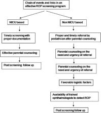
          The main barriers to early screening for ROP were related to availability of trained human resources, ignorance of “parents and health care personnel,” and distance from the point of care. This calls for training of ophthalmologists, advocacy with neonatologists and parents, and create systems for better coordination and compliance of the care providers.

          Related collections

          Most cited references13

          • Record: found
          • Abstract: found
          • Article: not found

          Aggressive posterior retinopathy of prematurity in large preterm babies in South India.

          To describe aggressive posterior retinopathy of prematurity (APROP) in a subset of premature babies, having gestational age (GA) of ≥28 weeks and birth weight (BW) of ≥1000 g. Retrospective observational case series. Case records of 99 babies, who were diagnosed to have APROP between July 2002 and October 2010 were reviewed. Fundus fluorescein angiography (FFA) was carried out in 19 babies. The mean GA was 31.7 weeks (range 28-35 weeks) and mean BW was 1572 g (range 1000-2310 g). All these babies received supplemental unblended oxygen 3 days or longer after birth. Of the 52 babies who had an eye exam in the neonatal intensive care unit prior to discharge, 35 babies had loss of vascularised retina from zone II to zone I and four babies from zone III to zone I, when examined as an outpatient. FFA revealed large geographic areas of vaso-obliteration (more than 30 disc areas) posterior to the shunt vessels within vascularised retina. Features of severe capillary bed loss in the vascularised retina were seen in our cases. Oxygen could be a precipitating factor in causing this retinopathy of prematurity in large babies.
            Bookmark
            • Record: found
            • Abstract: found
            • Article: not found

            Outcomes of a protocol-based management for zone 1 retinopathy of prematurity: the Indian Twin Cities ROP Screening Program report number 2.

            To report the outcomes of zone 1 aggressive posterior retinopathy of prematurity (ROP) treated with aggressive laser photocoagulation by a protocol-based approach over a 10-year period. Prospective, interventional case series. Tertiary eye institute and neonatal intensive care units in the twin cities of Hyderabad and Secunderabad in South India. Babies screened under the Indian Twin Cities Retinopathy of Prematurity Screening (ITCROPS) program between January 1, 1997 and March 31, 2007. Multiple sessions of laser photocoagulation at any sign of plus disease with abnormal shunt vessels with or without neovascularization in zone 1. Based on the anatomic status of the retina at the final follow-up, the outcome was classified into good (completely regressed with no vitreoretinal changes), fair (regression with vitreoretinal distortion), or poor (progression into partial or total retinal detachment involving macula). Descriptive statistics (mean and range) were used for gestational age at birth, birth weight, and post-conceptional age at first presentation and at the first intervention. Trends in incidence of zone 1 ROP over 1 decade were analyzed. Outcomes were expressed in terms of proportions and 95% confidence interval (CI). Of the 3654 babies screened, 227 eyes of 115 babies were detected to have zone 1 aggressive disease, defined as zone 1 retinal vessels having abnormal closed-loop shunts, dilation and tortuosity, flat new vessels, or rapid progression. Laser was performed in 169 eyes. Disease regressed with good outcome in 142 eyes (84%) (95% CI, 77.6-89.2) and progressed to poor outcome in 13 eyes (7.69%) (95% CI, 4.1-12.8), while fair outcome was seen in 6 eyes (3.55%) (95% CI, 1.3-7.5). Thus 148 babies (87.5%) (95% CI, 81.6-92.14) had a favorable outcome. The outcomes in 8 treated eyes (4.7%) lost to follow-up were unknown. Zone 1 aggressive ROP has a good outcome if the screening is done early; intervention is prompt and adequate, with frequent follow-up until complete regression is achieved. Copyright © 2011 Elsevier Inc. All rights reserved.
              Bookmark
              • Record: found
              • Abstract: found
              • Article: not found

              Outcome of universal newborn eye screening with wide-field digital retinal image acquisition system: a pilot study

              PurposeTo evaluate the outcome of universal newborn eye screening with wide-field digital retinal imaging (WFDRI) system.MethodsIn this pilot study, we examined 1152 apparently healthy newborn infants in the obstetrics and gynecology ward of a civil hospital in Eastern India over 1.5 years. The examination included external eye examination, red reflex test and fundus imaging by WFDRI (RetCam II, Clarity medical system, Pleasanton, CA, USA) by a trained optometrist. The pathologies detected, net monetary gain and skilled manpower saved were documented. The results were compared with three similar studies thus far published in the literature.ResultsOcular abnormality of any kind was seen in 172 (14.93%) babies. Retinal hemorrhage in 153 babies (88.9% of all abnormal findings) was the most common abnormality; it was bilateral in 118 (77.12%) babies and 4 babies had foveal hemorrhage. Other abnormalities included vitreous hemorrhage (n=1), congenital glaucoma (n=2), uveal coloboma (n=2), retinopathy mimicking retinopathy of prematurity (n=2), and cystic fovea (n=3). The retinal hemorrhages resolved spontaneously in all eyes. One baby with congenital glaucoma received surgery and the other was treated medically. The benefits included savings in skilled manpower, a net monetary gain of INR 4.195 million (US$ 62,612) and skilled manpower saving by 319.4 h.ConclusionsThe universal neonatal eye screening using WFDRI detected pathologies that needed immediate care or regular follow up; saved skilled manpower with a net monetary gain. But compared to a red reflex test the benefits were marginal in terms of detecting treatment warranting ocular pathologies.
                Bookmark

                Author and article information

                Journal
                Indian J Ophthalmol
                Indian J Ophthalmol
                IJO
                Indian Journal of Ophthalmology
                Wolters Kluwer - Medknow (India )
                0301-4738
                1998-3689
                June 2019
                : 67
                : 6
                : 824-827
                Affiliations
                [1]Miriam Hyman Children Eye Care Center, L V Prasad Eye Institute, Mithu Tulsi Chanrai Campus, Bhubaneswar, Odisha, India
                [1 ]Department of Ophthalmology, Tagore Hospital and Heart Center, Jalandhar City, Punjab, India
                [2 ]Srimati Kanuri Shantamma Centre for Vitreoretinal Disease, L V Prasad Eye Institute, Kallam Anji Reddy Campus, Hyderabad, Telangana, India
                Author notes
                Correspondence to: Dr. Tapas Ranjan Padhi, Vitreoretinal Services, L V Prasad Eye Institute, Mithu Tulsi Chanrai Campus, Patia, Bhubaneswar - 751 024, Odisha, India. E-mail: tapaspadhi254@ 123456gmail.com
                Article
                IJO-67-824
                10.4103/ijo.IJO_972_18
                6552581
                31124495
                b54aabeb-3ae9-4e62-8c38-0868ebaf505c
                Copyright: © 2019 Indian Journal of Ophthalmology

                This is an open access journal, and articles are distributed under the terms of the Creative Commons Attribution-NonCommercial-ShareAlike 4.0 License, which allows others to remix, tweak, and build upon the work non-commercially, as long as appropriate credit is given and the new creations are licensed under the identical terms.

                History
                : 09 June 2018
                : 10 January 2019
                Categories
                Original Article

                Ophthalmology & Optometry
                advanced retinopathy of prematurity,cicatricial retinopathy of prematurity,late presentation,retinopathy of prematurity,screening

                Comments

                Comment on this article