40
views
0
recommends
+1 Recommend
0 collections
    0
    shares
      • Record: found
      • Abstract: found
      • Article: found
      Is Open Access

      Cytoplasmic TAF2–TAF8–TAF10 complex provides evidence for nuclear holo–TFIID assembly from preformed submodules

      research-article

      Read this article at

      Bookmark
          There is no author summary for this article yet. Authors can add summaries to their articles on ScienceOpen to make them more accessible to a non-specialist audience.

          Abstract

          General transcription factor TFIID is a cornerstone of RNA polymerase II transcription initiation in eukaryotic cells. How human TFIID—a megadalton-sized multiprotein complex composed of the TATA-binding protein (TBP) and 13 TBP-associated factors (TAFs)—assembles into a functional transcription factor is poorly understood. Here we describe a heterotrimeric TFIID subcomplex consisting of the TAF2, TAF8 and TAF10 proteins, which assembles in the cytoplasm. Using native mass spectrometry, we define the interactions between the TAFs and uncover a central role for TAF8 in nucleating the complex. X-ray crystallography reveals a non-canonical arrangement of the TAF8–TAF10 histone fold domains. TAF2 binds to multiple motifs within the TAF8 C-terminal region, and these interactions dictate TAF2 incorporation into a core–TFIID complex that exists in the nucleus. Our results provide evidence for a stepwise assembly pathway of nuclear holo–TFIID, regulated by nuclear import of preformed cytoplasmic submodules.

          Abstract

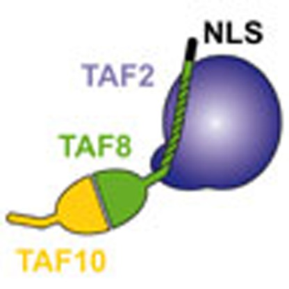
          TFIID is an essential transcription factor complex that controls the expression of most protein-coding genes in eukaryotes. Here the authors identify and characterize a complex containing TAF2, TAF8 and TAF10, which assembles in the cytoplasm before integration into the nuclear holo–TFIID complex.

          Related collections

          Most cited references55

          • Record: found
          • Abstract: found
          • Article: not found

          ESPript/ENDscript: Extracting and rendering sequence and 3D information from atomic structures of proteins.

          The fortran program ESPript was created in 1993, to display on a PostScript figure multiple sequence alignments adorned with secondary structure elements. A web server was made available in 1999 and ESPript has been linked to three major web tools: ProDom which identifies protein domains, PredictProtein which predicts secondary structure elements and NPS@ which runs sequence alignment programs. A web server named ENDscript was created in 2002 to facilitate the generation of ESPript figures containing a large amount of information. ENDscript uses programs such as BLAST, Clustal and PHYLODENDRON to work on protein sequences and such as DSSP, CNS and MOLSCRIPT to work on protein coordinates. It enables the creation, from a single Protein Data Bank identifier, of a multiple sequence alignment figure adorned with secondary structure elements of each sequence of known 3D structure. Similar 3D structures are superimposed in turn with the program PROFIT and a final figure is drawn with BOBSCRIPT, which shows sequence and structure conservation along the Calpha trace of the query. ESPript and ENDscript are available at http://genopole.toulouse.inra.fr/ESPript.
            Bookmark
            • Record: found
            • Abstract: found
            • Article: not found

            A high-resolution map of active promoters in the human genome.

            In eukaryotic cells, transcription of every protein-coding gene begins with the assembly of an RNA polymerase II preinitiation complex (PIC) on the promoter. The promoters, in conjunction with enhancers, silencers and insulators, define the combinatorial codes that specify gene expression patterns. Our ability to analyse the control logic encoded in the human genome is currently limited by a lack of accurate information regarding the promoters for most genes. Here we describe a genome-wide map of active promoters in human fibroblast cells, determined by experimentally locating the sites of PIC binding throughout the human genome. This map defines 10,567 active promoters corresponding to 6,763 known genes and at least 1,196 un-annotated transcriptional units. Features of the map suggest extensive use of multiple promoters by the human genes and widespread clustering of active promoters in the genome. In addition, examination of the genome-wide expression profile reveals four general classes of promoters that define the transcriptome of the cell. These results provide a global view of the functional relationships among transcriptional machinery, chromatin structure and gene expression in human cells.
              Bookmark
              • Record: found
              • Abstract: found
              • Article: not found

              Toward the structural genomics of complexes: crystal structure of a PE/PPE protein complex from Mycobacterium tuberculosis.

              The developing science called structural genomics has focused to date mainly on high-throughput expression of individual proteins, followed by their purification and structure determination. In contrast, the term structural biology is used to denote the determination of structures, often complexes of several macromolecules, that illuminate aspects of biological function. Here we bridge structural genomics to structural biology with a procedure for determining protein complexes of previously unknown function from any organism with a sequenced genome. From computational genomic analysis, we identify functionally linked proteins and verify their interaction in vitro by coexpression/copurification. We illustrate this procedure by the structural determination of a previously unknown complex between a PE and PPE protein from the Mycobacterium tuberculosis genome, members of protein families that constitute approximately 10% of the coding capacity of this genome. The predicted complex was readily expressed, purified, and crystallized, although we had previously failed in expressing individual PE and PPE proteins on their own. The reason for the failure is clear from the structure, which shows that the PE and PPE proteins mate along an extended apolar interface to form a four-alpha-helical bundle, where two of the alpha-helices are contributed by the PE protein and two by the PPE protein. Our entire procedure for the identification, characterization, and structural determination of protein complexes can be scaled to a genome-wide level.
                Bookmark

                Author and article information

                Journal
                Nat Commun
                Nat Commun
                Nature Communications
                Nature Pub. Group
                2041-1723
                14 January 2015
                : 6
                : 6011
                Affiliations
                [1 ]European Molecular Biology Laboratory, Grenoble Outstation , 6 rue Jules Horowitz, 38042 Grenoble, France
                [2 ]Unit for Virus Host-Cell Interactions, University Grenoble Alpes-EMBL-CNRS , 6 rue Jules Horowitz, 38042 Grenoble, France
                [3 ]Cellular Signaling and Nuclear Dynamics Program, Institut de Génétique et de Biologie Moléculaire et Cellulaire , UMR 7104, INSERM U964, 1 rue Laurent Fries, 67404 Illkirch, France
                [4 ]Proteomics Platform, Institut de Génétique et de Biologie Moléculaire et Cellulaire , UMR 7104, INSERM U964, 1 rue Laurent Fries, 67404 Illkirch, France
                [5 ]Integrated Structural Biology Department, Institut de Génétique et de Biologie Moléculaire et Cellulaire , UMR 7104, INSERM U964, 1 rue Laurent Fries, 67404 Illkirch, France
                [6 ]Chemistry Research Laboratory, University of Oxford , South Parks Road, Oxford OX1 3TA, UK
                [7 ]Wellcome Trust Centre for Cell Biology, University of Edinburgh , Mayfield Road, Edinburgh EH9 3JR, UK
                [8 ]Institute of Bioanalytics, Department of Biotechnology, Technische Universität Berlin , 13353 Berlin, Germany
                [9 ]School of Biochemistry, Bristol University , Bristol BS8 1TD, UK
                Author notes
                Article
                ncomms7011
                10.1038/ncomms7011
                4309443
                25586196
                b69db471-51ed-4ac4-b593-7d43d0035d2e
                Copyright © 2015, Nature Publishing Group, a division of Macmillan Publishers Limited. All Rights Reserved.

                This work is licensed under a Creative Commons Attribution 4.0 International License. The images or other third party material in this article are included in the article’s Creative Commons license, unless indicated otherwise in the credit line; if the material is not included under the Creative Commons license, users will need to obtain permission from the license holder to reproduce the material. To view a copy of this license, visit http://creativecommons.org/licenses/by/4.0/

                History
                : 13 August 2014
                : 02 December 2014
                Categories
                Article

                Uncategorized
                Uncategorized

                Comments

                Comment on this article