15
views
0
recommends
+1 Recommend
0 collections
    0
    shares
      • Record: found
      • Abstract: found
      • Article: found
      Is Open Access

      Cortical responses elicited by luminance and compound stimuli modulated by pseudo-random sequences: comparison between normal trichromats and congenital red-green color blinds

      research-article

      Read this article at

      Bookmark
          There is no author summary for this article yet. Authors can add summaries to their articles on ScienceOpen to make them more accessible to a non-specialist audience.

          Abstract

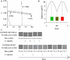
          Conventional pattern-reversal visual evoked cortical potential (VECP) shows positivity for luminance and chromatic equiluminant stimuli while conventional pattern-onset VECP shows positivity for luminance pattern-onset and negativity for chromatic pattern-onset. We evaluated how the presentation mode affects VECPs elicited by luminance and compound (luminance plus chromatic) pseudo-random stimulation. Eleven normal trichromats and 17 red-green color-blinds were studied. Pattern-reversal and pattern-onset luminance and compound (luminance plus red-green) gratings were temporally modulated by m-sequence. We used a cross-correlation routine to extract the first order kernel (K1) and the first and second slices of the second order kernel (K2.1 and K2.2, respectively) from the VECP response. We integrated the amplitude of VECP components as a function of time in order to estimate its magnitude for each stimulus condition. We also used a normalized cross-correlation method in order to test the similarity of the VECP components. The VECP components varied with the presentation mode and the presence of red-green contrast in the stimuli. In trichromats, for compound conditions, pattern-onset K1, K2.1, and K2.2, and pattern-reversal K2.1 and K2.2 had negative-dominated waveforms at 100 ms. Small negativity or small positivity were observed in dichromats. Trichromats had larger VECP magnitude than color-blinds for compound pattern-onset K1 (with large variability across subjects), compound pattern-onset and pattern-reversal K2.1, and compound pattern-reversal K2.2. Trichromats and color-blinds had similar VECP amplitude for compound pattern-reversal K1 and compound pattern-onset K2.2, as well as for all luminance conditions. The cross-correlation analysis showed high similarity between waveforms of compound pattern-onset K2.1 and pattern-reversal K2.2 as well as pattern-reversal K2.1 and K2.2. We suggest that compound pattern-reversal K2.1 is an appropriate response to study red-green color-opponent activity.

          Related collections

          Most cited references36

          • Record: found
          • Abstract: found
          • Article: not found

          ISCEV standard for clinical visual evoked potentials (2009 update).

          Visual evoked potentials (VEPs) can provide important diagnostic information regarding the functional integrity of the visual system. This document updates the ISCEV standard for clinical VEP testing and supersedes the 2004 standard. The major change in this revision is that test parameters have been made more precise to achieve better consistency of results within and between test centers. The ISCEV standard VEP protocols are defined for a single recording channel with a midline occipital active electrode. These protocols are intended for assessment of prechiasmal function; additional electrode sites are recommended for evaluation of chiasmal and postchiasmal function. ISCEV has selected a subset of stimulus and recording conditions that provide core clinical information and can be performed by most clinical electrophysiology laboratories throughout the world. These are: 1. Pattern-reversal VEPs elicited by checkerboard stimuli with large 1 degrees (i.e., 60 min of arc; min) and small 0.25 degrees (15 min) checks. 2. Pattern onset/offset VEPs elicited by checkerboard stimuli with large 1 degrees (60 min) and small 0.25 degrees (15 min) checks. 3. Flash VEP elicited by a brief luminance increment, a flash, which subtends a visual field of at least 20 degrees.
            Bookmark
            • Record: found
            • Abstract: found
            • Article: not found

            X and Y cells in the lateral geniculate nucleus of macaque monkeys.

            1. Cells of the lateral geniculate nucleus (l.g.n.) in macaque monkeys were sorted into two functional groups on the basis of spatial summation of visually evoked neural signals. 2. Cells were called X cells if their responses to contrast reversal of fine sine gratings were at the fundamental temporal modulation frequency with null positions one quarter of a cycle away from positions for peak response. Cells were called Y cells if their responses to such stimuli were at twice the modulation frequency and were approximately independent of spatial phase. 3. Ninety-nine percent of the cells in the four dorsal parvocellular layers of the l.g.n. were X cells; about seventy-five percent of the cells in the two ventral magnocellular layers were also X cells. The remainder were Y cells. 4. We confirmed previous findings that magnocellular cells had a shorter latency of response to electrical stimulation of the optic chiasm. 5. Magnocellular cells had much higher contrast sensitivities than did parvocellular cells. 6. Therefore, two distinct classes of X cells exist in the macaque l.g.n.: parvocellular X cells and magnocellular X cells. The great difference in their properties suggests that they have different functions in vision. The Y cells in the magnocellular layers form a third functional group with spatial properties distinctly different from the X cells. 7. We propose that the magnocellular layers of the macaque monkey's l.g.n. may be homologous to the A and A1 layers of the cat's l.g.n.
              Bookmark
              • Record: found
              • Abstract: found
              • Article: not found

              Identification, classification and anatomical segregation of cells with X-like and Y-like properties in the lateral geniculate nucleus of old-world primates.

              1. All the cells (158) that we studied in the lateral geniculate nuclei of Macaca nemestrina and Macaca irus could be distinguished as either X-like or Y-like on the basis of their responses to tests developed to classify cat retinal and lateral geniculate nucleus cells. These tests include responses to stationary spots, fast moving wands and moving gratings. 2. Response latencies to electrical stimulation of the optic chiasm were determined for 130 cells; no X-like cell showed a latency shorter than 1-7 ms, no Y-like cell showed a latency longer than 1-6 ms. Primate lateral geniculate nucleus cells with X-like properties thus receive their excitatory input from retinal cells with slowly conducting axons and these most probably include the tonic ganglion cells described by Gouras (1968, 1969); Y-like lateral geniculate nucleus cells are driven by retinal cells with faster conducting axons, most probably including the phasic ganglion cells described by Gouras. 3. Wiesel & Hubel (1966) classified monkey lateral geniculate nucleus cells into four main types based on their receptive-field properties, as revealed by spectrally and spatially distinct stimuli. We find that all Type I and Type II cells show X-like properties; all type IV cells show Y-like properties. Type III consists of a subtype that show X-like properties, here termed Type IIIx, and a subtype that show Y-like properties, here termed Type IIIy. 4. The first cells encountered as the micro-electrode reached the lateral geniculate nucleus were always X-like. In some penetrations only X-like cells were encountered as the electrode moved downward through the lateral geniculate nucleus. In the remaining penetrations, after recording X-like cells through most of the lateral geniculate nucleus, Y-like cells were then encountered. No X-like cells were found below Y-like cells. thus these two classes of cells are anatomically segregated within the primate lateral geniculate nucleus. Electrode marking showed the borger between X-like and Y-like cells to correspond to the border between the paro- and magnocellular layers of the lateral geniculate nucleus. Thus X-like cells (i.e. Types I, II and IIIx) occur in the parvocellular layers, Y-like cells (i.e. Types IIIy and IV)in the magnocellular layers.
                Bookmark

                Author and article information

                Contributors
                Journal
                Front Psychol
                Front Psychol
                Front. Psychol.
                Frontiers in Psychology
                Frontiers Media S.A.
                1664-1078
                28 January 2015
                2015
                : 6
                : 53
                Affiliations
                [1] 1Instituto de Ciências Biológicas – Universidade Federal do Pará Belém, Brazil
                [2] 2Núcleo de Medicina Tropical – Universidade Federal do Pará Belém, Brazil
                [3] 3Universidade Ceuma São Luís, Brazil
                Author notes

                Edited by: Marcelo Fernandes Costa, Universidade de São Paulo, Brazil

                Reviewed by: Sophie Wuerger, University of Liverpool, UK; Thomas Wachtler, Ludwig-Maximilians-Universität München, Germany

                *Correspondence: Givago S. Souza, Núcleo de Medicina Tropical – Universidade Federal do Pará, Av. Generalíssimo Deodoro 92 Umarizal 66055-240, Belém, Pará, Brazil e-mail: givagosouza@ 123456ufpa.br

                This article was submitted to Perception Science, a section of the journal Frontiers in Psychology.

                Article
                10.3389/fpsyg.2015.00053
                4309178
                ba7174ab-e08e-491c-bbe7-36d1d5b1efa6
                Copyright © 2015 Risuenho, Miquilini, Lacerda, Silveira and Souza.

                This is an open-access article distributed under the terms of the Creative Commons Attribution License (CC BY). The use, distribution or reproduction in other forums is permitted, provided the original author(s) or licensor are credited and that the original publication in this journal is cited, in accordance with accepted academic practice. No use, distribution or reproduction is permitted which does not comply with these terms.

                History
                : 13 August 2014
                : 12 January 2015
                Page count
                Figures: 6, Tables: 0, Equations: 0, References: 37, Pages: 8, Words: 0
                Categories
                Psychology
                Original Research Article

                Clinical Psychology & Psychiatry
                evoked potential,pseudo-random vecp,pattern-onset vecp,pattern-reversal vecp,color vision,trichromacy,daltonism

                Comments

                Comment on this article