11
views
0
recommends
+1 Recommend
0 collections
    0
    shares
      • Record: found
      • Abstract: found
      • Article: not found

      Immune-relevant aspects of murine models of head and neck cancer

      research-article
      1 , 2 , 2 , 3
      Oncogene
      experimental models, immunology

      Read this article at

      ScienceOpenPublisherPMC
      Bookmark
          There is no author summary for this article yet. Authors can add summaries to their articles on ScienceOpen to make them more accessible to a non-specialist audience.

          Abstract

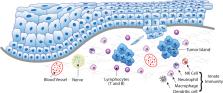
          Head and neck cancers (HNC) cause significant mortality and morbidity. There have been few advances in therapeutic management of HNC in the past 4 to 5 decades, which supports the need for studies focusing on HNC biology. In recent years, increased recognition of the relevance of the host response in cancer progression has led to novel therapeutic strategies and putative biomarkers of tumor aggressiveness. However, tumor-immune interactions are highly complex and vary with cancer type. Pre-clinical, in vivo models represent an important and necessary step in understanding biological processes involved in development, progression and treatment of HNC. Rodents (mice, rats, hamsters) are the most frequently used animal models in HNC research. The relevance and utility of information generated by studies in murine models is unquestionable, but it is also limited in application to tumor-immune interactions. In this review, we present information regarding the immune-specific characteristics of the murine models most commonly used in HNC research, including immunocompromised and immunocompetent animals. The particular characteristics of xenograft, chemically-induced, syngeneic, transgenic, and humanized models are discussed in order to provide context and insight for researchers interested in the in vivo study of tumor-immune interactions in HNC.

          Related collections

          Most cited references157

          • Record: found
          • Abstract: found
          • Article: not found

          The mechanism of double-strand DNA break repair by the nonhomologous DNA end-joining pathway.

          Double-strand DNA breaks are common events in eukaryotic cells, and there are two major pathways for repairing them: homologous recombination (HR) and nonhomologous DNA end joining (NHEJ). The various causes of double-strand breaks (DSBs) result in a diverse chemistry of DNA ends that must be repaired. Across NHEJ evolution, the enzymes of the NHEJ pathway exhibit a remarkable degree of structural tolerance in the range of DNA end substrate configurations upon which they can act. In vertebrate cells, the nuclease, DNA polymerases, and ligase of NHEJ are the most mechanistically flexible and multifunctional enzymes in each of their classes. Unlike repair pathways for more defined lesions, NHEJ repair enzymes act iteratively, act in any order, and can function independently of one another at each of the two DNA ends being joined. NHEJ is critical not only for the repair of pathologic DSBs as in chromosomal translocations, but also for the repair of physiologic DSBs created during variable (diversity) joining [V(D)J] recombination and class switch recombination (CSR). Therefore, patients lacking normal NHEJ are not only sensitive to ionizing radiation (IR), but also severely immunodeficient.
            Bookmark
            • Record: found
            • Abstract: found
            • Article: not found

            Inflammation: gearing the journey to cancer.

            Chronic inflammation plays a multifaceted role in carcinogenesis. Mounting evidence from preclinical and clinical studies suggests that persistent inflammation functions as a driving force in the journey to cancer. The possible mechanisms by which inflammation can contribute to carcinogenesis include induction of genomic instability, alterations in epigenetic events and subsequent inappropriate gene expression, enhanced proliferation of initiated cells, resistance to apoptosis, aggressive tumor neovascularization, invasion through tumor-associated basement membrane and metastasis, etc. Inflammation-induced reactive oxygen and nitrogen species cause damage to important cellular components (e.g., DNA, proteins and lipids), which can directly or indirectly contribute to malignant cell transformation. Overexpression, elevated secretion, or abnormal activation of proinflammatory mediators, such as cytokines, chemokines, cyclooxygenase-2, prostaglandins, inducible nitric oxide synthase, and nitric oxide, and a distinct network of intracellular signaling molecules including upstream kinases and transcription factors facilitate tumor promotion and progression. While inflammation promotes development of cancer, components of the tumor microenvironment, such as tumor cells, stromal cells in surrounding tissue and infiltrated inflammatory/immune cells generate an intratumoral inflammatory state by aberrant expression or activation of some proinflammatory molecules. Many of proinflammatory mediators, especially cytokines, chemokines and prostaglandins, turn on the angiogenic switches mainly controlled by vascular endothelial growth factor, thereby inducing inflammatory angiogenesis and tumor cell-stroma communication. This will end up with tumor angiogenesis, metastasis and invasion. Moreover, cellular microRNAs are emerging as a potential link between inflammation and cancer. The present article highlights the role of various proinflammatory mediators in carcinogenesis and their promise as potential targets for chemoprevention of inflammation-associated carcinogenesis.
              Bookmark
              • Record: found
              • Abstract: found
              • Article: not found

              IL-22, not simply a Th17 cytokine.

              Interleukin-22 (IL-22) has important functions in host defense at mucosal surfaces as well as in tissue repair. It is unique as a cytokine that is produced by immune cells, including T-helper (Th) cell subsets and innate lymphocytes, but acts only on non-hematopoietic stromal cells, in particular epithelial cells, keratinocytes, and hepatocytes. Although IL-22 is beneficial to the host in many infectious and inflammatory disorders, depending on the target tissue it can be pathogenic due to its inherent pro-inflammatory properties, which are further enhanced when IL-22 is released together with other pro-inflammatory cytokines, in particular IL-17. To avoid pathology, IL-22 and IL-17 production have to be controlled tightly and independently. While common factors such as signal transducer and activator of transcription 3 (STAT3) and retinoid orphan receptor γt (RORγt) drive the expression of both cytokines, other factors, such as c-Maf act specifically on IL-22 and enable the separate expression of either cytokine. Here, we discuss the production of IL-22 from various T-cell populations as well as protective versus pathogenic roles of IL-22. Finally, we focus on recent advances in our understanding of the molecular regulation of IL-22 in T cells. © 2013 John Wiley & Sons A/S. Published by Blackwell Publishing Ltd.
                Bookmark

                Author and article information

                Journal
                8711562
                6325
                Oncogene
                Oncogene
                Oncogene
                0950-9232
                1476-5594
                23 December 2018
                29 January 2019
                May 2019
                29 July 2019
                : 38
                : 21
                : 3973-3988
                Affiliations
                [1 ]Department of Diagnosis and Surgery, School of Dentistry at Araraquara, UNESP- Univ Estadual Paulista, Araraquara, SP, Brazil, University of Michigan, Ann Arbor, 48109, USA.
                [2 ]Periodontics and Oral Medicine, School of Dentistry, University of Michigan, Ann Arbor, 48109, USA.
                [3 ]Pathology, Medical School; University of Michigan, Ann Arbor, 48109, USA.
                Author notes
                Correspondence should be addressed to: Dr. Carlos Rossa Jr, UNESP- Univ Estadual Paulista, School of Dentistry in Araraquara, Department of Diagnosis and Surgery, CEP 14801-903, Araraquara, SP, Brazil. c.rossa@ 123456unesp.br
                Dr. Nisha J D’Silva, Department of Periodontics and Oral Medicine, University of Michigan School of Dentistry, 1011 North University Ave, Room G018, Ann Arbor, MI 48109-1078, USA, Phone: (734) 764-1543, Fax: (734) 764-2469, njdsilva@ 123456umich.edu
                Article
                NIHMS1516139
                10.1038/s41388-019-0686-9
                6533118
                30696955
                bb36f835-2ad1-45b6-80bb-61989618fc4d

                Users may view, print, copy, and download text and data-mine the content in such documents, for the purposes of academic research, subject always to the full Conditions of use: http://www.nature.com/authors/editorial_policies/license.html#terms

                History
                Categories
                Article

                Oncology & Radiotherapy
                experimental models,immunology
                Oncology & Radiotherapy
                experimental models, immunology

                Comments

                Comment on this article