35
views
0
recommends
+1 Recommend
0 collections
    5
    shares
      • Record: found
      • Abstract: found
      • Article: found
      Is Open Access

      Interactions between Amyloid-β and Hemoglobin: Implications for Amyloid Plaque Formation in Alzheimer's Disease

      research-article

      Read this article at

      Bookmark
          There is no author summary for this article yet. Authors can add summaries to their articles on ScienceOpen to make them more accessible to a non-specialist audience.

          Abstract

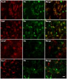
          Accumulation of amyloid-β (Aβ) peptides in the brain is one of the central pathogenic events in Alzheimer's disease (AD). However, why and how Aβ aggregates within the brain of AD patients remains elusive. Previously, we demonstrated hemoglobin (Hb) binds to Aβ and co-localizes with the plaque and vascular amyloid deposits in post-mortem AD brains. In this study, we further characterize the interactions between Hb and Aβ in vitro and in vivo and report the following observations: 1) the binding of Hb to Aβ required iron-containing heme; 2) other heme-containing proteins, such as myoglobin and cytochrome C, also bound to Aβ; 3) hemin-induced cytotoxicity was reduced in neuroblastoma cells by low levels of Aβ; 4) Hb was detected in neurons and glial cells of post-mortem AD brains and was up-regulated in aging and APP/PS1 transgenic mice; 5) microinjection of human Hb into the dorsal hippocampi of the APP/PS1 transgenic mice induced the formation of an envelope-like structure composed of Aβ surrounding the Hb droplets. Our results reveal an enhanced endogenous expression of Hb in aging brain cells, probably serving as a compensatory mechanism against hypoxia. In addition, Aβ binds to Hb and other hemoproteins via the iron-containing heme moiety, thereby reducing Hb/heme/iron-induced cytotoxicity. As some of the brain Hb could be derived from the peripheral circulation due to a compromised blood-brain barrier frequently observed in aged and AD brains, our work also suggests the genesis of some plaques may be a consequence of sustained amyloid accretion at sites of vascular injury.

          Related collections

          Most cited references59

          • Record: found
          • Abstract: found
          • Article: not found

          Common structure of soluble amyloid oligomers implies common mechanism of pathogenesis.

          Soluble oligomers are common to most amyloids and may represent the primary toxic species of amyloids, like the Abeta peptide in Alzheimer's disease (AD). Here we show that all of the soluble oligomers tested display a common conformation-dependent structure that is unique to soluble oligomers regardless of sequence. The in vitro toxicity of soluble oligomers is inhibited by oligomer-specific antibody. Soluble oligomers have a unique distribution in human AD brain that is distinct from fibrillar amyloid. These results indicate that different types of soluble amyloid oligomers have a common structure and suggest they share a common mechanism of toxicity.
            Bookmark
            • Record: found
            • Abstract: found
            • Article: not found

            Gene regulation and DNA damage in the ageing human brain.

            The ageing of the human brain is a cause of cognitive decline in the elderly and the major risk factor for Alzheimer's disease. The time in life when brain ageing begins is undefined. Here we show that transcriptional profiling of the human frontal cortex from individuals ranging from 26 to 106 years of age defines a set of genes with reduced expression after age 40. These genes play central roles in synaptic plasticity, vesicular transport and mitochondrial function. This is followed by induction of stress response, antioxidant and DNA repair genes. DNA damage is markedly increased in the promoters of genes with reduced expression in the aged cortex. Moreover, these gene promoters are selectively damaged by oxidative stress in cultured human neurons, and show reduced base-excision DNA repair. Thus, DNA damage may reduce the expression of selectively vulnerable genes involved in learning, memory and neuronal survival, initiating a programme of brain ageing that starts early in adult life.
              Bookmark
              • Record: found
              • Abstract: found
              • Article: not found

              Cerebral microvascular pathology in aging and Alzheimer's disease.

              The aging of the central nervous system and the development of incapacitating neurological diseases like Alzheimer's disease (AD) are generally associated with a wide range of histological and pathophysiological changes eventually leading to a compromised cognitive status. Although the diverse triggers of the neurodegenerative processes and their interactions are still the topic of extensive debate, the possible contribution of cerebrovascular deficiencies has been vigorously promoted in recent years. Various forms of cerebrovascular insufficiency such as reduced blood supply to the brain or disrupted microvascular integrity in cortical regions may occupy an initiating or intermediate position in the chain of events ending with cognitive failure. When, for example, vasoconstriction takes over a dominating role in the cerebral vessels, the perfusion rate of the brain can considerably decrease causing directly or through structural vascular damage a drop in cerebral glucose utilization. Consequently, cerebral metabolism can suffer a setback leading to neuronal damage and a concomitant suboptimal cognitive capacity. The present review focuses on the microvascular aspects of neurodegenerative processes in aging and AD with special attention to cerebral blood flow, neural metabolic changes and the abnormalities in microvascular ultrastructure. In this context, a few of the specific triggers leading to the prominent cerebrovascular pathology, as well as the potential neurological outcome of the compromised cerebral microvascular system are also going to be touched upon to a certain extent, without aiming at total comprehensiveness. Finally, a set of animal models are going to be presented that are frequently used to uncover the functional relationship between cerebrovascular factors and the damage to neural networks.
                Bookmark

                Author and article information

                Contributors
                Role: Editor
                Journal
                PLoS One
                plos
                plosone
                PLoS ONE
                Public Library of Science (San Francisco, USA )
                1932-6203
                2012
                6 March 2012
                : 7
                : 3
                : e33120
                Affiliations
                [1 ]Institute of Basic Medical Sciences, National Cheng Kung University, Tainan, Taiwan
                [2 ]Division of Neuroscience and Neuropathology, The School of Chinese Medicine for Post-Baccalaureate, I-Shou University, Kaohsiung, Taiwan
                [3 ]Institute of Behavioral Medicine, National Cheng Kung University, Tainan, Taiwan
                [4 ]Department of Cell Biology and Anatomy, National Cheng Kung University, Tainan, Taiwan
                Massachusetts General Hospital/Harvard Medical School, United States of America
                Author notes

                Conceived and designed the experiments: LY YK. Performed the experiments: JC CL YS TY. Analyzed the data: JC CL YS TY LY YK. Contributed reagents/materials/analysis tools: TY LY YK. Wrote the paper: JC LY YK.

                Article
                PONE-D-11-20767
                10.1371/journal.pone.0033120
                3295782
                22412990
                bc3af18f-55b1-4823-b7ed-851a4bba995b
                Chuang et al. This is an open-access article distributed under the terms of the Creative Commons Attribution License, which permits unrestricted use, distribution, and reproduction in any medium, provided the original author and source are credited.
                History
                : 19 October 2011
                : 10 February 2012
                Page count
                Pages: 10
                Categories
                Research Article
                Biology
                Biochemistry
                Cytochemistry
                Proteins
                Neuroscience
                Medicine
                Cardiovascular
                Neurology
                Dementia

                Uncategorized
                Uncategorized

                Comments

                Comment on this article