12
views
0
recommends
+1 Recommend
0 collections
    0
    shares
      • Record: found
      • Abstract: found
      • Article: found
      Is Open Access

      Catamenial Epilepsy: Discovery of an Extrasynaptic Molecular Mechanism for Targeted Therapy

      review-article

      Read this article at

      Bookmark
          There is no author summary for this article yet. Authors can add summaries to their articles on ScienceOpen to make them more accessible to a non-specialist audience.

          Abstract

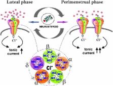
          Catamenial epilepsy is a type of refractory epilepsy characterized by seizure clusters around perimenstrual or periovulatory period. The pathophysiology of catamenial epilepsy still remains unclear, yet there are few animal models to study this gender-specific disorder. The pathophysiology of perimenstrual catamenial epilepsy involves the withdrawal of the progesterone-derived GABAergic neurosteroids due to the decline in progesterone level at the time of menstruation. These manifestations can be faithfully reproduced in rodents by specific neuroendocrine manipulations. Since mice and rats, like humans, have ovarian cycles with circulating hormones, they appear to be suitable animal models for studies of perimenstrual seizures. Recently, we created specific experimental models to mimic perimenstrual seizures. Studies in rat and mouse models of catamenial epilepsy show enhanced susceptibility to seizures or increased seizure exacerbations following neurosteroid withdrawal. During such a seizure exacerbation period, there is a striking decrease in the anticonvulsant effect of commonly prescribed antiepileptics, such as benzodiazepines, but an increase in the anticonvulsant potency of exogenous neurosteroids. We discovered an extrasynaptic molecular mechanism of catamenial epilepsy. In essence, extrasynaptic δGABA-A receptors are upregulated during perimenstrual-like neuroendocrine milieu. Consequently, there is enhanced antiseizure efficacy of neurosteroids in catamenial models because δGABA-A receptors confer neurosteroid sensitivity and greater seizure protection. Molecular mechanisms such as these offer a strong rationale for the clinical development of a neurosteroid replacement therapy for catamenial epilepsy.

          Related collections

          Most cited references79

          • Record: found
          • Abstract: found
          • Article: not found

          Ovarian cycle-linked changes in GABA(A) receptors mediating tonic inhibition alter seizure susceptibility and anxiety.

          Disturbances of neuronal excitability changes during the ovarian cycle may elevate seizure frequency in women with catamenial epilepsy and enhance anxiety in premenstrual dysphoric disorder (PMDD). The mechanisms underlying these changes are unknown, but they could result from the effects of fluctuations in progesterone-derived neurosteroids on the brain. Neurosteroids and some anxiolytics share an important site of action: tonic inhibition mediated by delta subunit-containing GABA(A) receptors (deltaGABA(A)Rs). Here we demonstrate periodic alterations in specific GABA(A)R subunits during the estrous cycle in mice, causing cyclic changes of tonic inhibition in hippocampal neurons. In late diestrus (high-progesterone phase), enhanced expression of deltaGABA(A)Rs increases tonic inhibition, and a reduced neuronal excitability is reflected by diminished seizure susceptibility and anxiety. Eliminating cycling of deltaGABA(A)Rs by antisense RNA treatment or gene knockout prevents the lowering of excitability during diestrus. Our findings are consistent with possible deficiencies in regulatory mechanisms controlling normal cycling of deltaGABA(A)Rs in individuals with catamenial epilepsy or PMDD.
            Bookmark
            • Record: found
            • Abstract: found
            • Article: not found

            Neurosteroids: endogenous role in the human brain and therapeutic potentials.

            This chapter provides an overview of neurosteroids, especially their impact on the brain, sex differences and their therapeutic potentials. Neurosteroids are synthesized within the brain and rapidly modulate neuronal excitability. They are classified as pregnane neurosteroids, such as allopregnanolone and allotetrahydrodeoxycorticosterone, androstane neurosteroids, such as androstanediol and etiocholanolone, and sulfated neurosteroids such as pregnenolone sulfate. Neurosteroids such as allopregnanolone are positive allosteric modulators of GABA-A receptors with powerful anti-seizure activity in diverse animal models. Neurosteroids increase both synaptic and tonic inhibition. They are endogenous regulators of seizure susceptibility, anxiety, and stress. Sulfated neurosteroids such as pregnenolone sulfate, which are negative GABA-A receptor modulators, are memory-enhancing agents. Sex differences in susceptibility to brain disorders could be due to neurosteroids and sexual dimorphism in specific structures of the human brain. Synthetic neurosteroids that exhibit better bioavailability and efficacy and drugs that enhance neurosteroid synthesis have therapeutic potential in anxiety, epilepsy, and other brain disorders. Clinical trials with the synthetic neurosteroid analog ganaxolone in the treatment of epilepsy have been encouraging. Neurosteroidogenic agents that lack benzodiazepine-like side effects show promise in the treatment of anxiety and depression. Copyright © 2010 Elsevier B.V. All rights reserved.
              Bookmark
              • Record: found
              • Abstract: found
              • Article: not found

              Characterization of brain neurons that express enzymes mediating neurosteroid biosynthesis.

              Allopregnanolone (ALLO) and tetrahydrodeoxycorticosterone (THDOC) are potent positive allosteric modulators of GABA action at GABA(A) receptors. ALLO and THDOC are synthesized in the brain from progesterone or deoxycorticosterone, respectively, by the sequential action of two enzymes: 5alpha-reductase (5alpha-R) type I and 3alpha-hydroxysteroid dehydrogenase (3alpha-HSD). This study evaluates 5alpha-R type I and 3alpha-HSD mRNA expression level in mouse brain by using in situ hybridization combined with glutamic acid decarboxylase 67/65, vesicular glutamate transporter 2, glial fibrillary acidic protein, and S100beta immunohistochemistry. We demonstrate that 5alpha-R type I and 3alpha-HSD colocalize in cortical, hippocampal, and olfactory bulb glutamatergic principal neurons and in some output neurons of the amygdala and thalamus. Neither 5alpha-R type I nor 3alpha-HSD mRNAs are expressed in S100beta- or glial fibrillary acidic protein-positive glial cells. Using glutamic acid decarboxylase 67/65 antibodies to mark GABAergic neurons, we failed to detect 5alpha-R type I and 3alpha-HSD in cortical and hippocampal GABAergic interneurons. However, 5alpha-R type I and 3alpha-HSD are significantly expressed in principal GABAergic output neurons, such as striatal medium spiny, reticular thalamic nucleus, and cerebellar Purkinje neurons. A similar distribution and cellular location of neurosteroidogenic enzymes was observed in rat brain. Taken together, these data suggest that ALLO and THDOC, which can be synthesized in principal output neurons, modulate GABA action at GABA(A) receptors, either with an autocrine or a paracrine mechanism or by reaching GABA(A) receptor intracellular sites through lateral membrane diffusion.
                Bookmark

                Author and article information

                Contributors
                Journal
                Front Cell Neurosci
                Front Cell Neurosci
                Front. Cell. Neurosci.
                Frontiers in Cellular Neuroscience
                Frontiers Media S.A.
                1662-5102
                22 April 2016
                2016
                : 10
                : 101
                Affiliations
                Department of Neuroscience and Experimental Therapeutics, Texas A&M University Health Science Center, College of Medicine Bryan, TX, USA
                Author notes

                Edited by: Rena Li, Roskamp Institute, USA

                Reviewed by: P. Hemachandra Reddy, Texas Tech University, USA; Thimmasettappa Thippeswamy, Iowa State University, USA

                *Correspondence: Doodipala Samba Reddy, reddy@ 123456medicine.tamhsc.edu
                Article
                10.3389/fncel.2016.00101
                4840555
                27147973
                bcb71e0a-ffb4-40d3-a0b8-7e6d158325d8
                Copyright © 2016 Reddy.

                This is an open-access article distributed under the terms of the Creative Commons Attribution License (CC BY). The use, distribution or reproduction in other forums is permitted, provided the original author(s) or licensor are credited and that the original publication in this journal is cited, in accordance with accepted academic practice. No use, distribution or reproduction is permitted which does not comply with these terms.

                History
                : 16 November 2015
                : 04 April 2016
                Page count
                Figures: 4, Tables: 1, Equations: 0, References: 85, Pages: 12, Words: 0
                Funding
                Funded by: National Institutes of Health 10.13039/100000002
                Award ID: NS052158 and NS051398
                Categories
                Neuroscience
                Review

                Neurosciences
                catamenial epilepsy,neurosteroid,gaba-a receptor,progesterone,mouse model
                Neurosciences
                catamenial epilepsy, neurosteroid, gaba-a receptor, progesterone, mouse model

                Comments

                Comment on this article