2
views
0
recommends
+1 Recommend
0 collections
    0
    shares
      • Record: found
      • Abstract: found
      • Article: found
      Is Open Access

      Protease‐activated receptor‐2 accelerates intestinal tumor formation through activation of nuclear factor‐κB signaling and tumor angiogenesis in Apc Min/+ mice

      research-article

      Read this article at

      Bookmark
          There is no author summary for this article yet. Authors can add summaries to their articles on ScienceOpen to make them more accessible to a non-specialist audience.

          Abstract

          Hepatocyte growth factor activator inhibitor‐1 (HAI‐1), encoded by the SPINT1 gene, is a membrane‐bound protease inhibitor expressed on the surface of epithelial cells. Hepatocyte growth factor activator inhibitor‐1 regulates type II transmembrane serine proteases that activate protease‐activated receptor‐2 (PAR‐2). We previously reported that deletion of Spint1 in Apc Min/+ mice resulted in accelerated formation of intestinal tumors, possibly through enhanced nuclear factor‐κB signaling. In this study, we examined the role of PAR‐2 in accelerating tumor formation in the Apc Min/+ model in the presence or absence of Spint1. We observed that knockout of the F2rl1 gene, encoding PAR‐2, not only eliminated the enhanced formation of intestinal tumors caused by Spint1 deletion, but also reduced tumor formation in the presence of Spint1. Exacerbation of anemia and weight loss associated with HAI‐1 deficiency was also normalized by compound deficiency of PAR‐2. Mechanistically, signaling triggered by deregulated protease activities increased nuclear translocation of RelA/p65, vascular endothelial growth factor expression, and vascular density in Apc Min/+‐induced intestinal tumors. These results suggest that serine proteases promote intestinal carcinogenesis through activation of PAR‐2, and that HAI‐1 plays a critical tumor suppressor role as an inhibitor of matriptase, kallikreins, and other PAR‐2 activating proteases.

          Abstract

          Hepatocyte growth factor activator inhibitor‐1 insufficiency induces protease‐activated receptor‐2 activation, resulting in tumor promotion through nuclear factor‐κB activation and tumor angiogenesis

          Related collections

          Most cited references56

          • Record: found
          • Abstract: found
          • Article: not found

          Thrombin signalling and protease-activated receptors.

          S Coughlin (2000)
          How does the coagulation protease thrombin regulate cellular behaviour? The protease-activated receptors (PARs) provide one answer. In concert with the coagulation cascade, these receptors provide an elegant mechanism linking mechanical information in the form of tissue injury or vascular leakage to cellular responses. Roles for PARs are beginning to emerge in haemostasis and thrombosis, inflammation, and perhaps even blood vessel development.
            Bookmark
            • Record: found
            • Abstract: found
            • Article: found
            Is Open Access

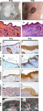
            Kallikrein 5 induces atopic dermatitis–like lesions through PAR2-mediated thymic stromal lymphopoietin expression in Netherton syndrome

            Netherton syndrome (NS) is a severe genetic skin disease with constant atopic manifestations that is caused by mutations in the serine protease inhibitor Kazal-type 5 (SPINK5) gene, which encodes the protease inhibitor lymphoepithelial Kazal-type–related inhibitor (LEKTI). Lack of LEKTI causes stratum corneum detachment secondary to epidermal proteases hyperactivity. This skin barrier defect favors allergen absorption and is generally regarded as the underlying cause for atopy in NS. We show for the first time that the pro-Th2 cytokine thymic stromal lymphopoietin (TSLP), the thymus and activation-regulated chemokine, and the macrophage-derived chemokine are overexpressed in LEKTI-deficient epidermis. This is part of an original biological cascade in which unregulated kallikrein (KLK) 5 directly activates proteinase-activated receptor 2 and induces nuclear factor κB–mediated overexpression of TSLP, intercellular adhesion molecule 1, tumor necrosis factor α, and IL8. This proinflammatory and proallergic pathway is independent of the primary epithelial failure and is activated under basal conditions in NS keratinocytes. This cell-autonomous process is already established in the epidermis of Spink5−/− embryos, and the resulting proinflammatory microenvironment leads to eosinophilic and mast cell infiltration in a skin graft model in nude mice. Collectively, these data establish that uncontrolled KLK5 activity in NS epidermis can trigger atopic dermatitis (AD)–like lesions, independently of the environment and the adaptive immune system. They illustrate the crucial role of protease signaling in skin inflammation and point to new therapeutic targets for NS as well as candidate genes for AD and atopy.
              Bookmark
              • Record: found
              • Abstract: found
              • Article: not found

              Role of tissue factor in cancer.

              Tissue factor (TF) is a transmembrane glycoprotein that localizes the coagulation serine protease factor VII/VIIa (FVII/VIIa) to the cell surface. The primary function of TF is to activate the clotting cascade. The TF:FVIIa complex also activates cells by cleavage of a G-protein coupled receptor called protease-activated receptor 2 (PAR2). TF is expressed by tumor cells and contributes to a variety of pathologic processes, such as thrombosis, metastasis, tumor growth, and tumor angiogenesis. For instance, tumor cells release TF-positive procoagulant microparticles into the circulation and these may trigger venous thromboembolism in patients with cancer. TF on circulating tumor cells also leads to the coating of the cells with fibrin that traps them within the microvasculature and facilitates hematogenous metastasis. In addition, TF:FVIIa-dependent activation of PAR2 on tumor cells increases tumor growth via an undefined mechanism. One possibility is that PAR2-dependent signaling increases the expression of proangiogenic proteins. Other studies have reported that endothelial cells in the tumor vasculature express TF and this may enhance angiogenesis. These results suggest that inhibition of TF should reduce several pathologic pathways that increase tumor growth and metastasis. This would represent a novel approach to anticancer therapy. Initial studies using inhibitors of the TF:FVIIa complex in mouse tumor models have produced encouraging results. Nevertheless, additional studies are needed to determine if this strategy can be successfully translated to the treatment of cancer patients.
                Bookmark

                Author and article information

                Contributors
                fukuchan@med.miyazaki-u.ac.jp
                Journal
                Cancer Sci
                Cancer Sci
                10.1111/(ISSN)1349-7006
                CAS
                Cancer Science
                John Wiley and Sons Inc. (Hoboken )
                1347-9032
                1349-7006
                24 February 2020
                April 2020
                : 111
                : 4 ( doiID: 10.1111/cas.v111.4 )
                : 1193-1202
                Affiliations
                [ 1 ] Department of Pathology University of Miyazaki Miyazaki Japan
                [ 2 ] Inserm U970 Paris Cardiovascular Research Center Université de Paris Paris France
                Author notes
                [*] [* ] Correspondence

                Tsuyoshi Fukushima, Section of Oncopathology and Regenerative Biology, Department of Pathology, Faculty of Medicine, University of Miyazaki, Miyazaki, Japan.

                Email: fukuchan@ 123456med.miyazaki-u.ac.jp

                Author information
                https://orcid.org/0000-0001-8210-5460
                https://orcid.org/0000-0001-9948-0451
                https://orcid.org/0000-0002-9350-777X
                Article
                CAS14335
                10.1111/cas.14335
                7156842
                31997435
                bd1c4bef-f8af-4ee8-8560-c0c8b15b4942
                © 2020 The Authors. Cancer Science published by John Wiley & Sons Australia, Ltd on behalf of Japanese Cancer Association.

                This is an open access article under the terms of the http://creativecommons.org/licenses/by-nc/4.0/ License, which permits use, distribution and reproduction in any medium, provided the original work is properly cited and is not used for commercial purposes.

                History
                : 11 September 2019
                : 19 January 2020
                : 23 January 2020
                Page count
                Figures: 6, Tables: 0, Pages: 10, Words: 6105
                Funding
                Funded by: Japan Society for the Promotion of Science KAKENHI , open-funder-registry 10.13039/501100001691;
                Award ID: 15K08311
                Award ID: 16H05175
                Award ID: 17K08764
                Funded by: Agence Nationale de la Recherche , open-funder-registry 10.13039/501100001665;
                Award ID: ANR‐15‐CE14‐0009
                Categories
                Original Article
                Original Articles
                Carcinogenesis
                Custom metadata
                2.0
                April 2020
                Converter:WILEY_ML3GV2_TO_JATSPMC version:5.8.0 mode:remove_FC converted:15.04.2020

                Oncology & Radiotherapy
                angiogenesis,colon cancer,hai‐1,par‐2,spint1
                Oncology & Radiotherapy
                angiogenesis, colon cancer, hai‐1, par‐2, spint1

                Comments

                Comment on this article