54
views
0
recommends
+1 Recommend
0 collections
    0
    shares
      • Record: found
      • Abstract: found
      • Article: found
      Is Open Access

      Identifying a Network of Brain Regions Involved in Aversion-Related Processing: A Cross-Species Translational Investigation

      review-article

      Read this article at

      Bookmark
          There is no author summary for this article yet. Authors can add summaries to their articles on ScienceOpen to make them more accessible to a non-specialist audience.

          Abstract

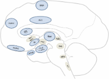
          The ability to detect and respond appropriately to aversive stimuli is essential for all organisms, from fruit flies to humans. This suggests the existence of a core neural network which mediates aversion-related processing. Human imaging studies on aversion have highlighted the involvement of various cortical regions, such as the prefrontal cortex, while animal studies have focused largely on subcortical regions like the periaqueductal gray and hypothalamus. However, whether and how these regions form a core neural network of aversion remains unclear. To help determine this, a translational cross-species investigation in humans (i.e., meta-analysis) and other animals (i.e., systematic review of functional neuroanatomy) was performed. Our results highlighted the recruitment of the anterior cingulate cortex, the anterior insula, and the amygdala as well as other subcortical (e.g., thalamus, midbrain) and cortical (e.g., orbitofrontal) regions in both animals and humans. Importantly, involvement of these regions remained independent of sensory modality. This study provides evidence for a core neural network mediating aversion in both animals and humans. This not only contributes to our understanding of the trans-species neural correlates of aversion but may also carry important implications for psychiatric disorders where abnormal aversive behavior can often be observed.

          Related collections

          Most cited references167

          • Record: found
          • Abstract: found
          • Article: not found

          Does rejection hurt? An FMRI study of social exclusion.

          A neuroimaging study examined the neural correlates of social exclusion and tested the hypothesis that the brain bases of social pain are similar to those of physical pain. Participants were scanned while playing a virtual ball-tossing game in which they were ultimately excluded. Paralleling results from physical pain studies, the anterior cingulate cortex (ACC) was more active during exclusion than during inclusion and correlated positively with self-reported distress. Right ventral prefrontal cortex (RVPFC) was active during exclusion and correlated negatively with self-reported distress. ACC changes mediated the RVPFC-distress correlation, suggesting that RVPFC regulates the distress of social exclusion by disrupting ACC activity.
            Bookmark
            • Record: found
            • Abstract: found
            • Article: not found

            Interoception: the sense of the physiological condition of the body.

            Converging evidence indicates that primates have a distinct cortical image of homeostatic afferent activity that reflects all aspects of the physiological condition of all tissues of the body. This interoceptive system, associated with autonomic motor control, is distinct from the exteroceptive system (cutaneous mechanoreception and proprioception) that guides somatic motor activity. The primary interoceptive representation in the dorsal posterior insula engenders distinct highly resolved feelings from the body that include pain, temperature, itch, sensual touch, muscular and visceral sensations, vasomotor activity, hunger, thirst, and 'air hunger'. In humans, a meta-representation of the primary interoceptive activity is engendered in the right anterior insula, which seems to provide the basis for the subjective image of the material self as a feeling (sentient) entity, that is, emotional awareness.
              Bookmark
              • Record: found
              • Abstract: found
              • Article: not found

              The reward circuit: linking primate anatomy and human imaging.

              Although cells in many brain regions respond to reward, the cortical-basal ganglia circuit is at the heart of the reward system. The key structures in this network are the anterior cingulate cortex, the orbital prefrontal cortex, the ventral striatum, the ventral pallidum, and the midbrain dopamine neurons. In addition, other structures, including the dorsal prefrontal cortex, amygdala, hippocampus, thalamus, and lateral habenular nucleus, and specific brainstem structures such as the pedunculopontine nucleus, and the raphe nucleus, are key components in regulating the reward circuit. Connectivity between these areas forms a complex neural network that mediates different aspects of reward processing. Advances in neuroimaging techniques allow better spatial and temporal resolution. These studies now demonstrate that human functional and structural imaging results map increasingly close to primate anatomy.
                Bookmark

                Author and article information

                Journal
                Front Integr Neurosci
                Front. Integr. Neurosci.
                Frontiers in Integrative Neuroscience
                Frontiers Research Foundation
                1662-5145
                07 August 2011
                03 October 2011
                2011
                : 5
                : 49
                Affiliations
                [1] 1simpleMind, Brain Imaging and Neuroethics Research Unit, Institute of Mental Health Research, University of Ottawa Ottawa, ON, Canada
                Author notes

                Edited by: John J. Foxe, Albert Einstein College of Medicine, USA

                Reviewed by: Rodrigo N. Romcy-Pereira, Federal University of Espírito Santo, Brazil; Janina Seubert, Monell Chemical Senses Center, USA

                *Correspondence: Dave J. Hayes, Mind, Brain Imaging and Neuroethics, Institute of Mental Health Research, Royal Ottawa Health Care Group, University of Ottawa, 1145 Carling Avenue, Room 6441, Ottawa, ON, Canada K1Z 7K4. e-mail: david.hayes@ 123456rohcg.on.ca
                Article
                10.3389/fnint.2011.00049
                3215229
                22102836
                bd4586fd-3d83-4182-9fc9-ddafbe2aa2d6
                Copyright © 2011 Hayes and Northoff.

                This is an open-access article subject to a non-exclusive license between the authors and Frontiers Media SA, which permits use, distribution and reproduction in other forums, provided the original authors and source are credited and other Frontiers conditions are complied with.

                History
                : 06 July 2011
                : 19 August 2011
                Page count
                Figures: 2, Tables: 5, Equations: 0, References: 207, Pages: 21, Words: 17314
                Categories
                Neuroscience
                Review Article

                Neurosciences
                animal models,aversion,meta-analysis,imaging,translational
                Neurosciences
                animal models, aversion, meta-analysis, imaging, translational

                Comments

                Comment on this article