574
views
0
recommends
+1 Recommend
0 collections
    0
    shares
      • Record: found
      • Abstract: found
      • Article: found
      Is Open Access

      Role of hypoxia-inducible factor-1α in preconditioning-induced protection of retinal ganglion cells in glaucoma

      research-article
      1 , 1 , 1 , 2 , 3 ,
      Molecular Vision
      Molecular Vision

      Read this article at

      Bookmark
          There is no author summary for this article yet. Authors can add summaries to their articles on ScienceOpen to make them more accessible to a non-specialist audience.

          Abstract

          Purpose

          We recently demonstrated in a mouse model of glaucoma that endogenous epigenetic mechanisms can be activated by a repetitive hypoxic preconditioning (RHP) stimulus to provide robust retinal ganglion cell (RGC) protection. Although we also provided evidence that RHP prevents or delays the apoptotic demise of the RGC soma, the mechanisms responsible for signaling this epigenetic response, as well as the effectors of the glaucoma-tolerant phenotype at the somatic and axonal levels, remain unidentified. In the present study, we used conditional mutant mice lacking hypoxia-inducible factor-1α (HIF-1α) in RGCs ( HIF-1α RGC-knockout [KO] mice) to test the hypothesis that RHP-mediated activation of this transcription factor in these cells protects them from glaucomatous injury.

          Methods

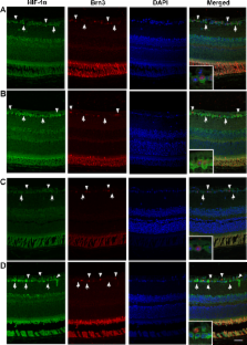
          Adult HIF-1α RGC-KO mice, generated by mating floxed HIF-1α mice with math5-Cre mice, were used. Experimental glaucoma was induced unilaterally in the HIF-1α RGC-KO mice and matched wild-types by elevating the intraocular pressure to 16–20 mmHg for 3 consecutive weeks, secondary to episcleral vein ligation. Mice of each genotype were randomized to either an RHP protocol (six total exposures to systemic hypoxia [11% oxygen], interspersed over a 2-week period, completed 3 days before ligation surgery) or to an untreated group. RGC soma and axon injury was quantified with Neuronal Nuclei (NeuN) immunohistochemistry in retinal flat mounts and SMI32 immunohistochemistry in cross sections of the post-laminar optic nerve, respectively.

          Results

          HIF-1α RGC-KO mice exhibited normal retinal function and morphology, and crosses of math5-Cre mice with floxed ROSA26 reporter mice confirmed Cre recombinase activity was confined to the RGC axons and soma. Untreated wild-type mice exhibited a 30±2% loss of RGC soma and a 31±3% loss of RGC axons after 3 weeks of intraocular hypertension (both p<0.05 versus fellow eye). The 90% and 81% improvement in soma and axon survival, respectively, observed in the wild-type mice treated with RHP (both p<0.05 versus the glaucoma eye in the untreated mice) was still observed to a near identical extent in the RHP-treated HIF-1α RGC-KO mice. RHP had no effect on the magnitude of intraocular pressure elevation in either the KO or wild-type groups, indicating that protection was realized in both genotypes in the face of ongoing intraocular hypertension.

          Conclusions

          These findings indicate that the robust, glaucomatous protection of the RGC soma and axons induced by RHP does not require HIF-1α-mediated transcription of survival genes and other adaptive responses within the RGCs themselves. Rather, we infer that RGC survival is augmented secondary to the activation of other hypoxia-sensitive transcription factors in RGCs and/or the action of diffusible HIF-1α target gene proteins released from neighboring retinal cells. Ideally, the involvement of such autocrine- and/or paracrine-based mechanisms would be confirmed in future studies, but distinct components of the integrated, pleiotropic, and multicellular basis of this endogenous epigenetic response may prove difficult to demonstrate experimentally, as we found in the present study.

          Related collections

          Most cited references41

          • Record: found
          • Abstract: found
          • Article: not found

          HIF-1 alpha is required for solid tumor formation and embryonic vascularization.

          The transcriptional response to lowered oxygen levels is mediated by the hypoxia-inducible transcription factor (HIF-1), a heterodimer consisting of the constitutively expressed aryl hydrocarbon receptor nuclear translocator (ARNT) and the hypoxic response factor HIF-1alpha. To study the role of the transcriptional hypoxic response in vivo we have targeted the murine HIF-1alpha gene. Loss of HIF-1alpha in embryonic stem (ES) cells dramatically retards solid tumor growth; this is correlated with a reduced capacity to release the angiogenic factor vascular endothelial growth factor (VEGF) during hypoxia. HIF-1alpha null mutant embryos exhibit clear morphological differences by embryonic day (E) 8.0, and by E8.5 there is a complete lack of cephalic vascularization, a reduction in the number of somites, abnormal neural fold formation and a greatly increased degree of hypoxia (measured by the nitroimidazole EF5). These data demonstrate the essential role of HIF-1alpha in controlling both embryonic and tumorigenic responses to variations in microenvironmental oxygenation.
            Bookmark
            • Record: found
            • Abstract: not found
            • Article: not found

            HIF1 and oxygen sensing in the brain.

              Bookmark
              • Record: found
              • Abstract: found
              • Article: not found

              Progressive ganglion cell degeneration precedes neuronal loss in a mouse model of glaucoma.

              Glaucoma is characterized by retinal ganglion cell (RGC) pathology and a progressive loss of vision. Previous studies suggest RGC death is responsible for vision loss in glaucoma, yet evidence from other neurodegenerative diseases suggests axonal degeneration, in the absence of neuronal loss, can significantly affect neuronal function. To characterize RGC degeneration in the DBA/2 mouse model of glaucoma, we quantified RGCs in mice of various ages using neuronal-specific nuclear protein (NeuN) immunolabeling, retrograde labeling, and optic nerve axon counts. Surprisingly, the number of NeuN-labeled RGCs did not decline significantly until 18 months of age, at which time a significant decrease in RGC somal size was also observed. Axon dysfunction and degeneration occurred before loss of NeuN-positive RGCs, because significant declines in RGC number assayed by retrograde tracers and axon counts were observed at 13 months. To examine whether axonal dysfunction/degeneration affected gene expression in RGC axons or somas, NeuN and neurofilament-heavy (NF-H) immunolabeling was performed along with quantitative reverse transcription-PCR for RGC-specific genes in retinas of aged DBA/2 mice. Although these mice had similar numbers of NeuN-positive RGCs, the expression of neurofilament light, Brn-3b, and Sncg mRNA varied; this variation in RGC-specific gene expression was correlated with the appearance of NF-H immunoreactive RGC axons. Together, these data support a progression of RGC degeneration in this model of glaucoma, beginning with loss of retrograde label, where axon dysfunction and degeneration precede neuronal loss. This progression of degeneration suggests a need to examine the RGC axon as a locus of pathology in glaucoma.
                Bookmark

                Author and article information

                Journal
                Mol Vis
                Mol. Vis
                MV
                Molecular Vision
                Molecular Vision
                1090-0535
                2013
                23 November 2013
                : 19
                : 2360-2372
                Affiliations
                [1 ]Department of Neurosurgery, Washington University School of Medicine St. Louis, MO
                [2 ]Department of Ophthalmology & Visual Sciences, Washington University School of Medicine St. Louis, MO
                [3 ]Department of and Cell Biology & Physiology, Washington University School of Medicine St. Louis, MO
                Author notes
                Correspondence to: Jeffrey M. Gidday, Department of Neurosurgery, Box 8057, Washington University School of Medicine, 660 S. Euclid Ave., 5th Floor McMillan, St. Louis, MO 63110; Phone: (314) 286-2795; FAX: (314) 286-2900; email: gidday@ 123456wustl.edu
                Article
                236 2013MOLVIS0353
                3850973
                24319330
                be1dc67c-bf9c-46e2-b32a-fe9eab87b0ea
                Copyright © 2013 Molecular Vision.

                This is an open-access article distributed under the terms of the Creative Commons Attribution License, which permits unrestricted use, distribution, and reproduction in any medium, provided the original work is properly cited, used for non-commercial purposes, and is not altered or transformed.

                History
                : 31 May 2013
                : 21 November 2013
                Categories
                Research Article
                Custom metadata
                Export to XML

                Vision sciences
                Vision sciences

                Comments

                Comment on this article