31
views
0
recommends
+1 Recommend
0 collections
    0
    shares
      • Record: found
      • Abstract: found
      • Article: found
      Is Open Access

      Cardiac Atrial Circadian Rhythms in PERIOD2::LUCIFERASE and per1:luc Mice: Amplitude and Phase Responses to Glucocorticoid Signaling and Medium Treatment

      research-article

      Read this article at

      Bookmark
          There is no author summary for this article yet. Authors can add summaries to their articles on ScienceOpen to make them more accessible to a non-specialist audience.

          Abstract

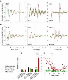
          Circadian rhythms in cardiac function are apparent in e.g., blood pressure, heart rate, and acute adverse cardiac events. A circadian clock in heart tissue has been identified, but entrainment pathways of this clock are still unclear. We cultured tissues of mice carrying bioluminescence reporters of the core clock genes, period 1 or 2 ( per1 luc or PER2 LUC) and compared in vitro responses of atrium to treatment with medium and a synthetic glucocorticoid (dexamethasone [DEX]) to that of the suprachiasmatic nucleus (SCN) and liver. We observed that PER2 LUC, but not per1 luc is rhythmic in atrial tissue, while both per1 luc and PER2 LUC exhibit rhythmicity in other cultured tissues. In contrast to the SCN and liver, both per1 luc and PER2 LUC bioluminescence amplitudes were increased in response to DEX treatment, and the PER2 LUC amplitude response was dependent on the time of treatment. Large phase-shift responses to both medium and DEX treatments were observed in the atrium, and phase responses to medium treatment were not attributed to serum content but the treatment procedure itself. The phase-response curves of atrium to both DEX and medium treatments were found to be different to the liver. Moreover, the time of day of the culturing procedure itself influenced the phase of the circadian clock in each of the cultured tissues, but the magnitude of this response was uniquely large in atrial tissue. The current data describe novel entrainment signals for the atrial circadian clock and specifically highlight entrainment by mechanical treatment, an intriguing observation considering the mechanical nature of cardiac tissue.

          Related collections

          Most cited references46

          • Record: found
          • Abstract: found
          • Article: not found

          Resetting of circadian time in peripheral tissues by glucocorticoid signaling.

          In mammals, circadian oscillators reside not only in the suprachiasmatic nucleus of the brain, which harbors the central pacemaker, but also in most peripheral tissues. Here, we show that the glucocorticoid hormone analog dexamethasone induces circadian gene expression in cultured rat-1 fibroblasts and transiently changes the phase of circadian gene expression in liver, kidney, and heart. However, dexamethasone does not affect cyclic gene expression in neurons of the suprachiasmatic nucleus. This enabled us to establish an apparent phase-shift response curve specifically for peripheral clocks in intact animals. In contrast to the central clock, circadian oscillators in peripheral tissues appear to remain responsive to phase resetting throughout the day.
            Bookmark
            • Record: found
            • Abstract: found
            • Article: not found

            Extensive and divergent circadian gene expression in liver and heart.

            Many mammalian peripheral tissues have circadian clocks; endogenous oscillators that generate transcriptional rhythms thought to be important for the daily timing of physiological processes. The extent of circadian gene regulation in peripheral tissues is unclear, and to what degree circadian regulation in different tissues involves common or specialized pathways is unknown. Here we report a comparative analysis of circadian gene expression in vivo in mouse liver and heart using oligonucleotide arrays representing 12,488 genes. We find that peripheral circadian gene regulation is extensive (> or = 8-10% of the genes expressed in each tissue), that the distributions of circadian phases in the two tissues are markedly different, and that very few genes show circadian regulation in both tissues. This specificity of circadian regulation cannot be accounted for by tissue-specific gene expression. Despite this divergence, the clock-regulated genes in liver and heart participate in overlapping, extremely diverse processes. A core set of 37 genes with similar circadian regulation in both tissues includes candidates for new clock genes and output genes, and it contains genes responsive to circulating factors with circadian or diurnal rhythms.
              Bookmark
              • Record: found
              • Abstract: found
              • Article: not found

              Suprachiasmatic nucleus: cell autonomy and network properties.

              The suprachiasmatic nucleus (SCN) is the primary circadian pacemaker in mammals. Individual SCN neurons in dispersed culture can generate independent circadian oscillations of clock gene expression and neuronal firing. However, SCN rhythmicity depends on sufficient membrane depolarization and levels of intracellular calcium and cAMP. In the intact SCN, cellular oscillations are synchronized and reinforced by rhythmic synaptic input from other cells, resulting in a reproducible topographic pattern of distinct phases and amplitudes specified by SCN circuit organization. The SCN network synchronizes its component cellular oscillators, reinforces their oscillations, responds to light input by altering their phase distribution, increases their robustness to genetic perturbations, and enhances their precision. Thus, even though individual SCN neurons can be cell-autonomous circadian oscillators, neuronal network properties are integral to normal function of the SCN.
                Bookmark

                Author and article information

                Contributors
                Role: Editor
                Journal
                PLoS One
                PLoS ONE
                plos
                plosone
                PLoS ONE
                Public Library of Science (San Francisco, USA )
                1932-6203
                2012
                23 October 2012
                : 7
                : 10
                : e47692
                Affiliations
                [1 ]Department of Biological Sciences, University of Notre Dame, Notre Dame, Indiana, United States of America
                [2 ]Department of Biochemistry and Physiology, Faculty of Health and Medical Sciences, University of Surrey, Guildford, Surrey, United Kingdom
                [3 ]Department of Physiology, Nankai University School of Medicine, Tianjin, People’s Republic of China
                Pennsylvania State University, United States of America
                Author notes

                Competing Interests: The authors have declared that no competing interests exist.

                Conceived and designed the experiments: DRV LL GED. Performed the experiments: DRV JS YX. Analyzed the data: DRV JS YX GED. Wrote the paper: DRV GED.

                Article
                PONE-D-12-18485
                10.1371/journal.pone.0047692
                3479129
                23110090
                c0d8075b-4bb5-4f94-8171-bdff39fc0ef9
                Copyright @ 2012

                This is an open-access article distributed under the terms of the Creative Commons Attribution License, which permits unrestricted use, distribution, and reproduction in any medium, provided the original author and source are credited.

                History
                : 25 June 2012
                : 17 September 2012
                Page count
                Pages: 13
                Funding
                The work was supported from grants to GED from the American Heart Association (Scientist Development Grant 10SDG4030011) and the National Institute of General Medical Sciences (R01-GM087508), and R01-GM083336. The funders had no role in study design, data collection and analysis, decision to publish, or preparation of the manuscript.
                Categories
                Research Article
                Biology
                Anatomy and Physiology
                Physiological Processes
                Chronobiology
                Cardiovascular System
                Genetics
                Gene Expression
                Molecular Genetics
                Model Organisms
                Animal Models
                Molecular Cell Biology
                Gene Expression
                Zoology
                Animal Physiology
                Medicine
                Anatomy and Physiology
                Cardiovascular System
                Cell Physiology
                Integrative Physiology
                Cardiovascular
                Cardiovascular Imaging
                Endocrinology
                Endocrine Physiology

                Uncategorized
                Uncategorized

                Comments

                Comment on this article