8
views
0
recommends
+1 Recommend
0 collections
    0
    shares
      • Record: found
      • Abstract: found
      • Article: found
      Is Open Access

      Platelet-Rich Plasma for Sport-Active Patients with Knee Osteoarthritis: Limited Return to Sport

      research-article

      Read this article at

      Bookmark
          There is no author summary for this article yet. Authors can add summaries to their articles on ScienceOpen to make them more accessible to a non-specialist audience.

          Abstract

          Objective

          To evaluate a cohort of sport-active patients suffering from cartilage degeneration and OA, in terms of clinical outcome and return to sport (RTS) after platelet-rich plasma (PRP) injective treatment.

          Design

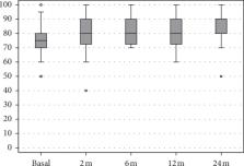
          This study included forty-seven sport-active patients ≤50 years old with unilateral symptomatic knee cartilage degeneration or OA. Patients received 3 PRP injections and were prospectively evaluated at baseline and then at 2, 6, 12, and 24 months follow-up by IKDC subjective EQ-VAS, and Tegner scores. Furthermore, patients were asked about their RTS, in terms of return to any sport level or to their activity level before symptoms onset.

          Results

          IKDC subjective score improved significantly at all follow-ups, changing from 59.2 ± 13.6 to 70.6 ± 13 at 12 months and to 76.7 ± 12.5 at 24 months ( p < 0.0005). A similar outcome was observed with the EQ-VAS score. Tegner score improved from 3.6 ± 1.4 to 4.8 ± 0.9 at 24 months ( p < 0.0005). A similar outcome was observed with the EQ-VAS score. Tegner score improved from 3.6 ± 1.4 to 4.8 ± 0.9 at 24 months ( p < 0.0005). A similar outcome was observed with the EQ-VAS score. Tegner score improved from 3.6 ± 1.4 to 4.8 ± 0.9 at 24 months ( p < 0.0005). A similar outcome was observed with the EQ-VAS score. Tegner score improved from 3.6 ± 1.4 to 4.8 ± 0.9 at 24 months (

          Conclusions

          Sport-active patients affected by knee OA can benefit from PRP injections, with pain and function improvement over time. However, results are less satisfactory in terms of RTS since only half can achieve the same sport level as before the onset of symptoms. Patients undergoing PRP treatment should be made aware of their low chances to go back to high-impact sport activities.

          Related collections

          Most cited references31

          • Record: found
          • Abstract: found
          • Article: not found

          Risk factors for the incidence and progression of radiographic knee osteoarthritis.

          Preventive strategies against knee osteoarthritis (OA) require a knowledge of risk factors that influence the initiation of the disorder and its subsequent progression. This population-based longitudinal study was performed to address this issue. Ninety-nine men and 255 women aged > or =55 years had baseline interviews and weight-bearing knee radiographs in 1990-1991. Repeat radiographs were obtained in 1995-1996 (mean followup duration 5.1 years, median age at followup 75.8 years). Risk factors assessed at baseline were tested for their association with incident and progressive radiographic knee OA by logistic regression. Rates of incidence and progression were 2.5% and 3.6% per year, respectively. After adjusting for age and sex, the risk of incident radiographic knee OA was significantly increased among subjects with higher baseline body mass index (odds ratio [OR] 18.3, 95% confidence interval [95% CI] 5.1-65.1, highest versus lowest third), previous knee injury (OR 4.8, 95% CI 1.0-24.1), and a history of regular sports participation (OR 3.2, 95% CI 1.1-9.1). Knee pain at baseline (OR 2.4, 95% CI 0.7-8.0) and Heberden's nodes (OR 2.0, 95% CI 0.7-5.7) were weakly associated with progression. Analyses based on individual radiographic features (osteophyte formation and joint space narrowing) supported differences in risk factors for either feature. Most currently recognized risk factors for prevalent knee OA (obesity, knee injury, and physical activity) influence incidence more than radiographic progression. Furthermore, these factors might selectively influence osteophyte formation more than joint space narrowing. These findings are consistent with knee OA being initiated by joint injury, but with progression being a consequence of impaired intrinsic repair capacity.
            Bookmark
            • Record: found
            • Abstract: found
            • Article: not found

            Patient satisfaction after knee arthroplasty: a report on 27,372 knees operated on between 1981 and 1995 in Sweden.

            During a validation process of the Swedish Knee Arthroplasty Register (SKAR), living registered patients were sent a questionnaire to ask if they had been reoperated on. This gave an opportunity to pose a simple four-point question with respect to patient satisfaction which 95% of patients answered. We analyzed the answers of patients operated on between 1981 and 1995 and found that only 8% of the patients were dissatisfied regarding their knee arthroplasty 2-17 years postoperatively. The satisfaction rate was constant, regardless of when the operation had been performed during the 15-year period. The proportion of satisfied patients was affected by the preoperative diagnosis, patients operated on for a long-standing disease more often being satisfied than those with a short disease-duration. There was no difference in proportions of satisfied patients, whether they had primarily been operated on with a total knee arthroplasty (TKA) or a medial unicompartmental arthroplasty (UKA). For TKAs performed with primary patellar resurfacing, there was a higher ratio of satisfied patients than for TKAs not resurfaced, but this increased ratio diminished with time passed since the primary operation. Unrevised knees had a higher proportion of satisfied patients than knees that had been subject to revision, and among patients revised for medial UKA, the proportion of satisfied patients was higher than among patients revised for TKA. We conclude that satisfaction after knee arthroplasty is stable and long-lasting in unrevised cases and that even after revision most patients are satisfied.
              Bookmark
              • Record: found
              • Abstract: found
              • Article: not found

              Autologous platelets as a source of proteins for healing and tissue regeneration

              Summary Platelets are known for their role in haemostasis where they help prevent blood loss at sites of vascular injury. To do this, they adhere, aggregate and form a procoagulant surface leading to thrombin generation and fibrin formation. Platelets also release substances that promote tissue repair and influence the reactivity of vascular and other blood cells in angiogenesis and inflammation. They contain storage pools of growth factors including PDGF, TGF-β and VEGF as well as cytokines including proteins such as PF4 and CD40L. Chemokines and newly synthesised active metabolites are also released. The fact that platelets secrete growth factors and active metabolites means that their applied use can have a positive influence in clinical situations requiring rapid healing and tissue regeneration. Their administration in fibrin clot or fibrin glue provides an adhesive support that can confine secretion to a chosen site. Additionally, the presentation of growth factors attached to platelets and/or fibrin may result in enhanced activity over recombinant proteins. Dental implant surgery with guided bone regeneration is one situation where an autologous platelet-rich clot clearly accelerates ossification after tooth extraction and/or around titanium implants. The end result is both marked reductions in the time required for implant stabilisation and an improved success rate. Orthopaedic surgery, muscle and/or tendon repair, reversal of skin ulcers, hole repair in eye surgery and cosmetic surgery are other situations where autologous platelets accelerate healing. Our aim is to review these advances and discuss the ways in which platelets may provide such unexpected beneficial therapeutic effects.
                Bookmark

                Author and article information

                Contributors
                Journal
                Biomed Res Int
                Biomed Res Int
                BMRI
                BioMed Research International
                Hindawi
                2314-6133
                2314-6141
                2020
                31 January 2020
                : 2020
                : 8243865
                Affiliations
                1Clinica Ortopedica e Traumatologica II IRCCS Istituto Ortopedico Rizzoli, Bologna, Italy
                2Servizio Trasfusionale Unico Metropolitano, Bologna, Italy
                3Applied and Translational Research (ATR) Center IRCCS Istituto Ortopedico Rizzoli, Bologna, Italy
                Author notes

                Academic Editor: Willeke F. Daamen

                Author information
                https://orcid.org/0000-0001-7066-4038
                https://orcid.org/0000-0002-2411-7508
                https://orcid.org/0000-0001-6352-9671
                https://orcid.org/0000-0002-8136-0977
                Article
                10.1155/2020/8243865
                7013341
                32076616
                c1b75d07-e9c6-48d1-8ea4-702a08bcd61c
                Copyright © 2020 Sante Alessandro Altamura et al.

                This is an open access article distributed under the Creative Commons Attribution License, which permits unrestricted use, distribution, and reproduction in any medium, provided the original work is properly cited.

                History
                : 6 June 2019
                : 25 November 2019
                : 18 December 2019
                Categories
                Clinical Study

                Comments

                Comment on this article