28
views
0
recommends
+1 Recommend
0 collections
    0
    shares
      • Record: found
      • Abstract: found
      • Article: found
      Is Open Access

      Colocalization of Tectal Inputs With Amygdala-Projecting Neurons in the Macaque Pulvinar

      research-article

      Read this article at

      Bookmark
          There is no author summary for this article yet. Authors can add summaries to their articles on ScienceOpen to make them more accessible to a non-specialist audience.

          Abstract

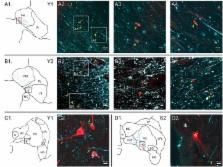
          Neuropsychological and neuroimaging studies have suggested the presence of a fast, subcortical route for the processing of emotionally-salient visual information in the primate brain. This putative pathway consists of the superior colliculus (SC), pulvinar and amygdala. While the presence of such a pathway has been confirmed in sub-primate species, it has yet to be documented in the primate brain using conventional anatomical methods. We injected retrograde tracers into the amygdala and anterograde tracers into the colliculus, and examined regions of colocalization of these signals within the pulvinar of the macaque. Anterograde tracers injected into the SC labeled axonal projections within the pulvinar, primarily within the oral, lateral and medial subdivisions. These axonal projections from the colliculus colocalized with cell bodies within the pulvinar that were labeled by retrograde tracer injected into the lateral amygdala. This zone of overlap was most notable in the medial portions of the medial (PM), oral (PO) and inferior pulvinar (PI), and was often densely concentrated in the vicinity of the brachium of the SC. These data provide an anatomical basis for the previously suggested pathway mediating fast processing of emotionally salient information.

          Related collections

          Most cited references66

          • Record: found
          • Abstract: found
          • Article: not found

          Masked presentations of emotional facial expressions modulate amygdala activity without explicit knowledge.

          Functional magnetic resonance imaging (fMRI) of the human brain was used to study whether the amygdala is activated in response to emotional stimuli, even in the absence of explicit knowledge that such stimuli were presented. Pictures of human faces bearing fearful or happy expressions were presented to 10 normal, healthy subjects by using a backward masking procedure that resulted in 8 of 10 subjects reporting that they had not seen these facial expressions. The backward masking procedure consisted of 33 msec presentations of fearful or happy facial expressions, their offset coincident with the onset of 167 msec presentations of neutral facial expressions. Although subjects reported seeing only neutral faces, blood oxygen level-dependent (BOLD) fMRI signal in the amygdala was significantly higher during viewing of masked fearful faces than during the viewing of masked happy faces. This difference was composed of significant signal increases in the amygdala to masked fearful faces as well as significant signal decreases to masked happy faces, consistent with the notion that the level of amygdala activation is affected differentially by the emotional valence of external stimuli. In addition, these facial expressions activated the sublenticular substantia innominata (SI), where signal increases were observed to both fearful and happy faces--suggesting a spatial dissociation of territories that respond to emotional valence versus salience or arousal value. This study, using fMRI in conjunction with masked stimulus presentations, represents an initial step toward determining the role of the amygdala in nonconscious processing.
            Bookmark
            • Record: found
            • Abstract: found
            • Article: not found

            Structure and function of visual area MT.

            The small visual area known as MT or V5 has played a major role in our understanding of the primate cerebral cortex. This area has been historically important in the concept of cortical processing streams and the idea that different visual areas constitute highly specialized representations of visual information. MT has also proven to be a fertile culture dish--full of direction- and disparity-selective neurons--exploited by many labs to study the neural circuits underlying computations of motion and depth and to examine the relationship between neural activity and perception. Here we attempt a synthetic overview of the rich literature on MT with the goal of answering the question, What does MT do?
              Bookmark
              • Record: found
              • Abstract: found
              • Article: not found

              The mammalian superior colliculus: laminar structure and connections.

              The superior colliculus is a laminated midbrain structure that acts as one of the centers organizing gaze movements. This review will concentrate on sensory and motor inputs to the superior colliculus, on its internal circuitry, and on its connections with other brainstem gaze centers, as well as its extensive outputs to those structures with which it is reciprocally connected. This will be done in the context of its laminar arrangement. Specifically, the superficial layers receive direct retinal input, and are primarily visual sensory in nature. They project upon the visual thalamus and pretectum to influence visual perception. These visual layers also project upon the deeper layers, which are both multimodal, and premotor in nature. Thus, the deep layers receive input from both somatosensory and auditory sources, as well as from the basal ganglia and cerebellum. Sensory, association, and motor areas of cerebral cortex provide another major source of collicular input, particularly in more encephalized species. For example, visual sensory cortex terminates superficially, while the eye fields target the deeper layers. The deeper layers are themselves the source of a major projection by way of the predorsal bundle which contributes collicular target information to the brainstem structures containing gaze-related burst neurons, and the spinal cord and medullary reticular formation regions that produce head turning.
                Bookmark

                Author and article information

                Contributors
                Journal
                Front Neural Circuits
                Front Neural Circuits
                Front. Neural Circuits
                Frontiers in Neural Circuits
                Frontiers Media S.A.
                1662-5110
                24 October 2018
                2018
                : 12
                : 91
                Affiliations
                [1] 1Interdisciplinary Program in Neuroscience, Georgetown University School of Medicine , Washington, DC, United States
                [2] 2Department of Pharmacology and Physiology, Georgetown University School of Medicine , Washington, DC, United States
                [3] 3Department of Neuroscience, Georgetown University School of Medicine , Washington, DC, United States
                [4] 4Laboratory of Neuropsychology, National Institute of Mental Health (NIMH) , Bethesda, ML, United States
                Author notes

                Edited by: Tadashi Isa, Kyoto University, Japan

                Reviewed by: Hisao Nishijo, University of Toyama, Japan; Paul J. May, University of Mississippi Medical Center, United States

                *Correspondence: Ludise Malkova malkoval@ 123456georgetown.edu
                Article
                10.3389/fncir.2018.00091
                6207581
                30405362
                c2478abb-1a2f-446f-a9fd-ae5635b029b5
                Copyright © 2018 Elorette, Forcelli, Saunders and Malkova.

                This is an open-access article distributed under the terms of the Creative Commons Attribution License (CC BY). The use, distribution or reproduction in other forums is permitted, provided the original author(s) and the copyright owner(s) are credited and that the original publication in this journal is cited, in accordance with accepted academic practice. No use, distribution or reproduction is permitted which does not comply with these terms.

                History
                : 21 June 2018
                : 03 October 2018
                Page count
                Figures: 7, Tables: 2, Equations: 0, References: 71, Pages: 12, Words: 8676
                Categories
                Neuroscience
                Original Research

                Neurosciences
                subcortical,superior colliculus,blindsight,retrograde,anterograde,anatomy
                Neurosciences
                subcortical, superior colliculus, blindsight, retrograde, anterograde, anatomy

                Comments

                Comment on this article