46
views
0
recommends
+1 Recommend
0 collections
    0
    shares
      • Record: found
      • Abstract: found
      • Article: found
      Is Open Access

      Deep learning for chest radiograph diagnosis: A retrospective comparison of the CheXNeXt algorithm to practicing radiologists

      research-article

      Read this article at

      Bookmark
          There is no author summary for this article yet. Authors can add summaries to their articles on ScienceOpen to make them more accessible to a non-specialist audience.

          Abstract

          Background

          Chest radiograph interpretation is critical for the detection of thoracic diseases, including tuberculosis and lung cancer, which affect millions of people worldwide each year. This time-consuming task typically requires expert radiologists to read the images, leading to fatigue-based diagnostic error and lack of diagnostic expertise in areas of the world where radiologists are not available. Recently, deep learning approaches have been able to achieve expert-level performance in medical image interpretation tasks, powered by large network architectures and fueled by the emergence of large labeled datasets. The purpose of this study is to investigate the performance of a deep learning algorithm on the detection of pathologies in chest radiographs compared with practicing radiologists.

          Methods and findings

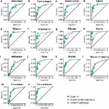
          We developed CheXNeXt, a convolutional neural network to concurrently detect the presence of 14 different pathologies, including pneumonia, pleural effusion, pulmonary masses, and nodules in frontal-view chest radiographs. CheXNeXt was trained and internally validated on the ChestX-ray8 dataset, with a held-out validation set consisting of 420 images, sampled to contain at least 50 cases of each of the original pathology labels. On this validation set, the majority vote of a panel of 3 board-certified cardiothoracic specialist radiologists served as reference standard. We compared CheXNeXt’s discriminative performance on the validation set to the performance of 9 radiologists using the area under the receiver operating characteristic curve (AUC). The radiologists included 6 board-certified radiologists (average experience 12 years, range 4–28 years) and 3 senior radiology residents, from 3 academic institutions. We found that CheXNeXt achieved radiologist-level performance on 11 pathologies and did not achieve radiologist-level performance on 3 pathologies. The radiologists achieved statistically significantly higher AUC performance on cardiomegaly, emphysema, and hiatal hernia, with AUCs of 0.888 (95% confidence interval [CI] 0.863–0.910), 0.911 (95% CI 0.866–0.947), and 0.985 (95% CI 0.974–0.991), respectively, whereas CheXNeXt’s AUCs were 0.831 (95% CI 0.790–0.870), 0.704 (95% CI 0.567–0.833), and 0.851 (95% CI 0.785–0.909), respectively. CheXNeXt performed better than radiologists in detecting atelectasis, with an AUC of 0.862 (95% CI 0.825–0.895), statistically significantly higher than radiologists' AUC of 0.808 (95% CI 0.777–0.838); there were no statistically significant differences in AUCs for the other 10 pathologies. The average time to interpret the 420 images in the validation set was substantially longer for the radiologists (240 minutes) than for CheXNeXt (1.5 minutes). The main limitations of our study are that neither CheXNeXt nor the radiologists were permitted to use patient history or review prior examinations and that evaluation was limited to a dataset from a single institution.

          Conclusions

          In this study, we developed and validated a deep learning algorithm that classified clinically important abnormalities in chest radiographs at a performance level comparable to practicing radiologists. Once tested prospectively in clinical settings, the algorithm could have the potential to expand patient access to chest radiograph diagnostics.

          Abstract

          In their study, Pranav Rajpurkar and colleagues test a deep learning algorithm that classifies clinically important abnormalities in chest radiographs.

          Author summary

          Why was this study done?
          • Chest radiographs are the most common medical imaging test in the world and critical for diagnosing common thoracic diseases.

          • Radiograph interpretation is a time-consuming task, and there is shortage of qualified trained radiologists in many healthcare systems.

          • Deep learning algorithms that have been developed to provide diagnostic chest radiograph interpretation have not been compared to expert human radiologist performance.

          What did the researchers do and find?
          • We developed a deep learning algorithm to concurrently detect 14 clinically important pathologies in chest radiographs.

          • The algorithm can also localize parts of the image most indicative of each pathology.

          • We evaluated the algorithm against 9 practicing radiologists on a validation set of 420 images for which the majority vote of 3 cardiothoracic specialty radiologists served as ground truth.

          • The algorithm achieved performance equivalent to the practicing radiologists on 10 pathologies, better on 1 pathology, and worse on 3 pathologies.

          • Radiologists labeled the 420 images in 240 minutes on average, and the algorithm labeled them in 1.5 minutes.

          What do these findings mean?
          • Deep learning algorithms can diagnose certain pathologies in chest radiographs at a level comparable to practicing radiologists on a single institution dataset.

          • After clinical validation, algorithms such as the one presented in this work could be used to increase access to rapid, high-quality chest radiograph interpretation.

          Related collections

          Most cited references32

          • Record: found
          • Abstract: not found
          • Conference Proceedings: not found

          Learning Deep Features for Discriminative Localization

            Bookmark
            • Record: found
            • Abstract: found
            • Article: not found

            Training and Validating a Deep Convolutional Neural Network for Computer-Aided Detection and Classification of Abnormalities on Frontal Chest Radiographs.

            Convolutional neural networks (CNNs) are a subtype of artificial neural network that have shown strong performance in computer vision tasks including image classification. To date, there has been limited application of CNNs to chest radiographs, the most frequently performed medical imaging study. We hypothesize CNNs can learn to classify frontal chest radiographs according to common findings from a sufficiently large data set.
              Bookmark
              • Record: found
              • Abstract: found
              • Article: not found

              A deep learning-based prediction model for gamma evaluation in patient-specific quality assurance.

              Patient-specific quality assurance (QA) measurement is conducted to confirm the accuracy of dose delivery. However, measurement is time-consuming and places a heavy workload on the medical physicists and radiological technologists. In this study, we proposed a prediction model for gamma evaluation, based on deep learning. We applied the model to a QA measurement dataset of prostate cancer cases to evaluate its practicality.
                Bookmark

                Author and article information

                Contributors
                Role: ConceptualizationRole: Data curationRole: Formal analysisRole: InvestigationRole: MethodologyRole: Project administrationRole: SoftwareRole: ValidationRole: VisualizationRole: Writing – original draftRole: Writing – review & editing
                Role: ConceptualizationRole: Data curationRole: Formal analysisRole: InvestigationRole: MethodologyRole: SoftwareRole: ValidationRole: VisualizationRole: Writing – original draftRole: Writing – review & editing
                Role: Formal analysisRole: InvestigationRole: MethodologyRole: SoftwareRole: ValidationRole: VisualizationRole: Writing – original draftRole: Writing – review & editing
                Role: Data curationRole: MethodologyRole: SoftwareRole: VisualizationRole: Writing – original draft
                Role: Data curationRole: MethodologyRole: SoftwareRole: VisualizationRole: Writing – original draft
                Role: Data curationRole: MethodologyRole: SoftwareRole: VisualizationRole: Writing – original draft
                Role: Data curationRole: MethodologyRole: SoftwareRole: VisualizationRole: Writing – original draft
                Role: Data curationRole: MethodologyRole: SoftwareRole: VisualizationRole: Writing – original draft
                Role: Data curationRole: MethodologyRole: SoftwareRole: VisualizationRole: Writing – original draft
                Role: ConceptualizationRole: SupervisionRole: Writing – review & editing
                Role: Data curationRole: InvestigationRole: Writing – review & editing
                Role: Data curationRole: Writing – review & editing
                Role: ConceptualizationRole: Data curationRole: Project administrationRole: Writing – original draftRole: Writing – review & editing
                Role: Data curationRole: Writing – review & editing
                Role: Data curationRole: Writing – review & editing
                Role: Data curationRole: Writing – review & editing
                Role: Data curationRole: Writing – review & editing
                Role: Data curationRole: Writing – review & editing
                Role: Data curationRole: Writing – review & editing
                Role: ConceptualizationRole: Project administrationRole: SupervisionRole: Writing – review & editing
                Role: ConceptualizationRole: Data curationRole: SupervisionRole: VisualizationRole: Writing – original draftRole: Writing – review & editing
                Role: Academic Editor
                Journal
                PLoS Med
                PLoS Med
                plos
                plosmed
                PLoS Medicine
                Public Library of Science (San Francisco, CA USA )
                1549-1277
                1549-1676
                20 November 2018
                November 2018
                : 15
                : 11
                : e1002686
                Affiliations
                [1 ] Department of Computer Science, Stanford University, Stanford, California, United States of America
                [2 ] Department of Medicine, Quantitative Sciences Unit, Stanford University, Stanford, California, United States of America
                [3 ] Department of Radiology, Stanford University, Stanford, California, United States of America
                [4 ] Department of Radiology, Duke University, Durham, North Carolina, United States of America
                [5 ] Department of Radiology, University of Colorado, Denver, Colorado, United States of America
                Edinburgh University, UNITED KINGDOM
                Author notes

                I have read the journal's policy and the authors of this manuscript have the following competing interests: CPL holds shares in whiterabbit.ai and Nines.ai, is on the Advisory Board of Nuance Communications and on the Board of Directors for the Radiological Society of North America, and has other research support from Philips, GE Healthcare, and Philips Healthcare. MPL holds shares in and serves on the Advisory Board for Nines.ai. None of these organizations have a financial interest in the results of this study.

                ‡These authors share first authorship on, and contributed equally to, this work.

                Author information
                http://orcid.org/0000-0002-8030-3727
                http://orcid.org/0000-0002-0395-4403
                http://orcid.org/0000-0002-8972-8051
                http://orcid.org/0000-0001-9860-3368
                http://orcid.org/0000-0003-2741-4046
                http://orcid.org/0000-0002-9354-9486
                http://orcid.org/0000-0003-1317-984X
                Article
                PMEDICINE-D-18-01880
                10.1371/journal.pmed.1002686
                6245676
                30457988
                c2c9d382-a39f-4da5-b185-759ca0f8d773
                © 2018 Rajpurkar et al

                This is an open access article distributed under the terms of the Creative Commons Attribution License, which permits unrestricted use, distribution, and reproduction in any medium, provided the original author and source are credited.

                History
                : 29 May 2018
                : 3 October 2018
                Page count
                Figures: 3, Tables: 1, Pages: 17
                Funding
                The authors received no specific funding for this work. This study was made possible via infrastructure support from the Stanford Center for Artificial Intelligence in Medicine and Imaging ( AIMI.stanford.edu).
                Categories
                Research Article
                People and Places
                Population Groupings
                Professions
                Medical Personnel
                Radiologists
                Physical Sciences
                Mathematics
                Applied Mathematics
                Algorithms
                Research and Analysis Methods
                Simulation and Modeling
                Algorithms
                Computer and Information Sciences
                Artificial Intelligence
                Machine Learning
                Deep Learning
                Medicine and Health Sciences
                Diagnostic Medicine
                Diagnostic Radiology
                Research and Analysis Methods
                Imaging Techniques
                Diagnostic Radiology
                Medicine and Health Sciences
                Radiology and Imaging
                Diagnostic Radiology
                Medicine and Health Sciences
                Diagnostic Medicine
                Cancer Detection and Diagnosis
                Medicine and Health Sciences
                Oncology
                Cancer Detection and Diagnosis
                Medicine and Health Sciences
                Oncology
                Cancers and Neoplasms
                Lung and Intrathoracic Tumors
                Medicine and Health Sciences
                Pulmonology
                Pneumonia
                Medicine and Health Sciences
                Diagnostic Medicine
                Signs and Symptoms
                Hernia
                Medicine and Health Sciences
                Pathology and Laboratory Medicine
                Signs and Symptoms
                Hernia
                Custom metadata
                The data used in this study is third party and is publicly hosted by the National Institutes of Health Clinical Center at https://nihcc.app.box.com/v/ChestXray-NIHCC. The test set annotations are not made publicly available to preserve the integrity of the test results when hosting public model evaluation. All other data is included in the paper, its Supporting Information files, and at the following Box link (which contains code as well): https://stanfordmedicine.box.com/s/b3gk9qnanzrdocqge0pbuh07mreu5x7y.

                Medicine
                Medicine

                Comments

                Comment on this article