23
views
0
recommends
+1 Recommend
1 collections
    0
    shares

      International Journal of Nanomedicine (submit here)

      This international, peer-reviewed Open Access journal by Dove Medical Press focuses on the application of nanotechnology in diagnostics, therapeutics, and drug delivery systems throughout the biomedical field. Sign up for email alerts here.

      105,621 Monthly downloads/views I 7.033 Impact Factor I 10.9 CiteScore I 1.22 Source Normalized Impact per Paper (SNIP) I 1.032 Scimago Journal & Country Rank (SJR)

      • Record: found
      • Abstract: found
      • Article: found
      Is Open Access

      Airspace Dimension Assessment with nanoparticles reflects lung density as quantified by MRI

      research-article

      Read this article at

      ScienceOpenPublisherPMC
      Bookmark
          There is no author summary for this article yet. Authors can add summaries to their articles on ScienceOpen to make them more accessible to a non-specialist audience.

          Abstract

          Background

          Airspace Dimension Assessment with inhaled nanoparticles is a novel method to determine distal airway morphology. This is the first empirical study using Airspace Dimension Assessment with nanoparticles (AiDA) to estimate distal airspace radius. The technology is relatively simple and potentially accessible in clinical outpatient settings.

          Method

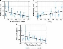
          Nineteen never-smoking volunteers performed nanoparticle inhalation tests at multiple breath-hold times, and the difference in nanoparticle concentration of inhaled and exhaled gas was measured. An exponential decay curve was fitted to the concentration of recovered nanoparticles, and airspace dimensions were assessed from the half-life of the decay. Pulmonary tissue density was measured using magnetic resonance imaging (MRI).

          Results

          The distal airspace radius measured by AiDA correlated with lung tissue density as measured by MRI ( ρ = −0.584; p = 0.0086). The linear intercept of the logarithm of the exponential decay curve correlated with forced expiratory volume in one second (FEV 1) ( ρ = 0.549; p = 0.0149).

          Conclusion

          The AiDA method shows potential to be developed into a tool to assess conditions involving changes in distal airways, eg, emphysema. The intercept may reflect airway properties; this finding should be further investigated.

          Most cited references17

          • Record: found
          • Abstract: found
          • Article: not found

          "Density mask". An objective method to quantitate emphysema using computed tomography.

          We used a computed tomography (CT) scanner program ("density mask") that highlights voxels within a given density range to quantitate emphysema by defining areas of abnormally low attenuation. We compared different density masks, mean lung attenuation, visual assessment of emphysema and the pathologic grade of emphysema in 28 patients undergoing lung resection for tumor. In each patient, a single representative CT image was compared with corresponding pathologic specimens of tissue. There was good correlation between the extent of emphysema as assessed by the density mask and the pathologic grade of emphysema. The optimal attenuation level to define areas of emphysema may vary in different scanners, but, once determined for a particular scanner, the density mask accurately assesses the extent of emphysema and eliminates interobserver and intraobserver variability. It has the added advantage of determining the exact percentage of lung parenchyma showing changes consistent with emphysema.
            Bookmark
            • Record: found
            • Abstract: found
            • Article: not found

            Pulmonary emphysema: objective quantification at multi-detector row CT--comparison with macroscopic and microscopic morphometry.

            To prospectively compare pulmonary function tests and helical computed tomographic (CT) indexes for quantifying pulmonary emphysema with macroscopic and microscopic morphometry. The investigation was approved by the local ethics committee, and written informed consent was obtained from patients. Multi-detector row CT of the thorax was performed with simultaneous acquisition of four 1-mm sections in 80 patients (57 men, 23 women; age range, 38-79 years) referred for surgical resection of lung cancer. From the raw data, 1.25-mm-thick sections were reconstructed at 10-mm intervals. Relative areas of lung with attenuation coefficients lower than nine thresholds and eight percentiles of the distribution of attenuation coefficients were calculated. Relative areas and percentiles were compared with areas found macroscopically to have emphysema and with two microscopic indexes assessed on resected specimens. Pulmonary function tests were measured 24-48 hours before surgery. Spearman correlation coefficients were calculated between each set of CT data obtained with the nine tested thresholds and eight percentiles with macroscopic and microscopic measurements. For relative lung areas, the strongest correlation with macroscopy was observed with a threshold of -970 HU (r = 0.543, P < .001) and that with microscopy was observed at -960 and -970 HU, depending on the index considered (r = 0.592, P < .001 and r = -0.546, P < .001, respectively). For percentiles, 1st percentile showed the strongest correlation with both macroscopy (r = -0.463, P < .001) and microscopy (r = -0.573, P < .001; and r = 0.523, P < .001 for each microscopic measurement). Forced expiratory volume in 1 second and vital capacity ratio, diffusing capacity of lung for carbon monoxide, and each of the three CT indexes were complementary to predict microscopic indexes. Relative lung areas with attenuation coefficients lower than -960 or -970 HU and 1st percentile are valid indexes to quantify pulmonary emphysema on multi-detector row CT scans. Copyright RSNA, 2006.
              Bookmark
              • Record: found
              • Abstract: not found
              • Article: not found

              Measurement of pulmonary emphysema.

                Bookmark

                Author and article information

                Journal
                Int J Nanomedicine
                Int J Nanomedicine
                International Journal of Nanomedicine
                International Journal of Nanomedicine
                Dove Medical Press
                1176-9114
                1178-2013
                2018
                21 May 2018
                : 13
                : 2989-2995
                Affiliations
                [1 ]Department of Medical Imaging and Physiology, Skåne University Hospital, Malmö, Sweden
                [2 ]Department of Translational Medicine, Lund University, Malmö, Sweden
                [3 ]Department of Medical Radiation Physics, Lund University, Malmö, Sweden
                [4 ]Department of Design Sciences, Lund University, Lund, Sweden
                [5 ]Department of Hematology, Oncology and Radiation Physics, Skåne University Hospital, Malmö, Sweden
                Author notes
                Correspondence: H Laura Aaltonen, Email laura.aaltonen@ 123456med.lu.se
                [*]

                These authors contributed equally to this work

                Article
                ijn-13-2989
                10.2147/IJN.S160331
                5968779
                c4a4eb0e-4f62-4e6e-bac3-9c6e16b30e31
                © 2018 Aaltonen et al. This work is published and licensed by Dove Medical Press Limited

                The full terms of this license are available at https://www.dovepress.com/terms.php and incorporate the Creative Commons Attribution – Non Commercial (unported, v3.0) License ( http://creativecommons.org/licenses/by-nc/3.0/). By accessing the work you hereby accept the Terms. Non-commercial uses of the work are permitted without any further permission from Dove Medical Press Limited, provided the work is properly attributed.

                History
                Categories
                Original Research

                Molecular medicine
                nanoparticles,respiratory diagnostics,distal airspaces,airspace dimension assessment with nanoparticles,magnetic resonance densitometry

                Comments

                Comment on this article