16
views
0
recommends
+1 Recommend
0 collections
    0
    shares
      • Record: found
      • Abstract: found
      • Article: found
      Is Open Access

      KRIT1 Loss-Of-Function Associated with Cerebral Cavernous Malformation Disease Leads to Enhanced S-Glutathionylation of Distinct Structural and Regulatory Proteins

      research-article

      Read this article at

      Bookmark
          There is no author summary for this article yet. Authors can add summaries to their articles on ScienceOpen to make them more accessible to a non-specialist audience.

          Abstract

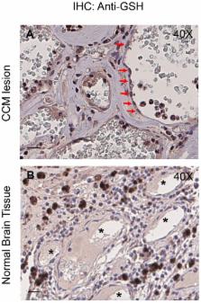
          Loss-of-function mutations in the KRIT1 gene are associated with the pathogenesis of cerebral cavernous malformations (CCMs), a major cerebrovascular disease still awaiting therapies. Accumulating evidence demonstrates that KRIT1 plays an important role in major redox-sensitive mechanisms, including transcriptional pathways and autophagy, which play major roles in cellular homeostasis and defense against oxidative stress, raising the possibility that KRIT1 loss has pleiotropic effects on multiple redox-sensitive systems. Using previously established cellular models, we found that KRIT1 loss-of-function affects the glutathione (GSH) redox system, causing a significant decrease in total GSH levels and increase in oxidized glutathione disulfide (GSSG), with a consequent deficit in the GSH/GSSG redox ratio and GSH-mediated antioxidant capacity. Redox proteomic analyses showed that these effects are associated with increased S-glutathionylation of distinct proteins involved in adaptive responses to oxidative stress, including redox-sensitive chaperonins, metabolic enzymes, and cytoskeletal proteins, suggesting a novel molecular signature of KRIT1 loss-of-function. Besides providing further insights into the emerging pleiotropic functions of KRIT1, these findings point definitively to KRIT1 as a major player in redox biology, shedding new light on the mechanistic relationship between KRIT1 loss-of-function and enhanced cell sensitivity to oxidative stress, which may eventually lead to cellular dysfunctions and CCM disease pathogenesis.

          Related collections

          Most cited references76

          • Record: found
          • Abstract: found
          • Article: not found

          Glutathione catalysis and the reaction mechanisms of glutathione-dependent enzymes.

          Glutathione-dependent catalysis is a metabolic adaptation to chemical challenges encountered by all life forms. In the course of evolution, nature optimized numerous mechanisms to use glutathione as the most versatile nucleophile for the conversion of a plethora of sulfur-, oxygen- or carbon-containing electrophilic substances. This comprehensive review summarizes fundamental principles of glutathione catalysis and compares the structures and mechanisms of glutathione-dependent enzymes, including glutathione reductase, glutaredoxins, glutathione peroxidases, peroxiredoxins, glyoxalases 1 and 2, glutathione transferases and MAPEG. Moreover, open mechanistic questions, evolutionary aspects and the physiological relevance of glutathione catalysis are discussed for each enzyme family. It is surprising how little is known about many glutathione-dependent enzymes, how often reaction geometries and acid-base catalysts are neglected, and how many mechanistic puzzles remain unsolved despite almost a century of research. On the one hand, several enzyme families with non-related protein folds recognize the glutathione moiety of their substrates. On the other hand, the thioredoxin fold is often used for glutathione catalysis. Ancient as well as recent structural changes of this fold did not only significantly alter the reaction mechanism, but also resulted in completely different protein functions. Glutathione-dependent enzymes are excellent study objects for structure-function relationships and molecular evolution. Notably, in times of systems biology, the outcome of models on glutathione metabolism and redox regulation is more than questionable as long as fundamental enzyme properties are neither studied nor understood. Furthermore, several of the presented mechanisms could have implications for drug development. This article is part of a Special Issue entitled Cellular functions of glutathione. Copyright © 2012 Elsevier B.V. All rights reserved.
            Bookmark
            • Record: found
            • Abstract: found
            • Article: not found

            Glutathione dysregulation and the etiology and progression of human diseases.

            Glutathione (GSH) plays an important role in a multitude of cellular processes, including cell differentiation, proliferation, and apoptosis, and as a result, disturbances in GSH homeostasis are implicated in the etiology and/or progression of a number of human diseases, including cancer, diseases of aging, cystic fibrosis, and cardiovascular, inflammatory, immune, metabolic, and neurodegenerative diseases. Owing to the pleiotropic effects of GSH on cell functions, it has been quite difficult to define the role of GSH in the onset and/or the expression of human diseases, although significant progress is being made. GSH levels, turnover rates, and/or oxidation state can be compromised by inherited or acquired defects in the enzymes, transporters, signaling molecules, or transcription factors that are involved in its homeostasis, or from exposure to reactive chemicals or metabolic intermediates. GSH deficiency or a decrease in the GSH/glutathione disulfide ratio manifests itself largely through an increased susceptibility to oxidative stress, and the resulting damage is thought to be involved in diseases, such as cancer, Parkinson's disease, and Alzheimer's disease. In addition, imbalances in GSH levels affect immune system function, and are thought to play a role in the aging process. Just as low intracellular GSH levels decrease cellular antioxidant capacity, elevated GSH levels generally increase antioxidant capacity and resistance to oxidative stress, and this is observed in many cancer cells. The higher GSH levels in some tumor cells are also typically associated with higher levels of GSH-related enzymes and transporters. Although neither the mechanism nor the implications of these changes are well defined, the high GSH content makes cancer cells chemoresistant, which is a major factor that limits drug treatment. The present report highlights and integrates the growing connections between imbalances in GSH homeostasis and a multitude of human diseases.
              Bookmark
              • Record: found
              • Abstract: found
              • Article: not found

              S-glutathionylation uncouples eNOS and regulates its cellular and vascular function.

              Endothelial nitric oxide synthase (eNOS) is critical in the regulation of vascular function, and can generate both nitric oxide (NO) and superoxide (O(2)(•-)), which are key mediators of cellular signalling. In the presence of Ca(2+)/calmodulin, eNOS produces NO, endothelial-derived relaxing factor, from l-arginine (l-Arg) by means of electron transfer from NADPH through a flavin containing reductase domain to oxygen bound at the haem of an oxygenase domain, which also contains binding sites for tetrahydrobiopterin (BH(4)) and l-Arg. In the absence of BH(4), NO synthesis is abrogated and instead O(2)(•-) is generated. While NOS dysfunction occurs in diseases with redox stress, BH(4) repletion only partly restores NOS activity and NOS-dependent vasodilation. This suggests that there is an as yet unidentified redox-regulated mechanism controlling NOS function. Protein thiols can undergo S-glutathionylation, a reversible protein modification involved in cellular signalling and adaptation. Under oxidative stress, S-glutathionylation occurs through thiol-disulphide exchange with oxidized glutathione or reaction of oxidant-induced protein thiyl radicals with reduced glutathione. Cysteine residues are critical for the maintenance of eNOS function; we therefore speculated that oxidative stress could alter eNOS activity through S-glutathionylation. Here we show that S-glutathionylation of eNOS reversibly decreases NOS activity with an increase in O(2)(•-) generation primarily from the reductase, in which two highly conserved cysteine residues are identified as sites of S-glutathionylation and found to be critical for redox-regulation of eNOS function. We show that eNOS S-glutathionylation in endothelial cells, with loss of NO and gain of O(2)(•-) generation, is associated with impaired endothelium-dependent vasodilation. In hypertensive vessels, eNOS S-glutathionylation is increased with impaired endothelium-dependent vasodilation that is restored by thiol-specific reducing agents, which reverse this S-glutathionylation. Thus, S-glutathionylation of eNOS is a pivotal switch providing redox regulation of cellular signalling, endothelial function and vascular tone.
                Bookmark

                Author and article information

                Journal
                Antioxidants (Basel)
                Antioxidants (Basel)
                antioxidants
                Antioxidants
                MDPI
                2076-3921
                17 January 2019
                January 2019
                : 8
                : 1
                : 27
                Affiliations
                [1 ]Department of Clinical Sciences (DISCO), Section of Biochemistry, Biology and Physics, Marche Polytechnic University, 60131 Ancona, Italy; l.cianfruglia@ 123456univpm.it (L.C.); principato@ 123456univpm.it (G.P.)
                [2 ]CCM Italia Research Network, National Coordination Center at the Department of Clinical and Biological Sciences, University of Turin, Orbassano, 10043 Torino, Italy; andrea.perrelli@ 123456unito.it (A.P.); claudia.fornelli@ 123456unito.it (C.F.); cinzia.antognelli@ 123456unipg.it (C.A.); francesca.retta@ 123456edu.unito.it (F.R.); valerio.benedetti@ 123456unito.it (V.B.); paola.cassoni@ 123456unito.it (P.C.)
                [3 ]Department of Clinical and Biological Sciences, University of Torino, Orbassano, 10043 Torino, Italy
                [4 ]Department of Chemistry, Biology and Biotechnology, University of Perugia, 06123 Perugia, Italy; alessandro.magini@ 123456unipg.it (A.M.); carla.emiliani@ 123456unipg.it (C.E.)
                [5 ]Department of Life and Environmental Sciences (DISVA), Marche Polytechnic University, 60131 Ancona, Italy; s.gorbi@ 123456univpm.it
                [6 ]Proteomics & Mass Spectrometry Laboratory, ISPAAM, National Research Council, 80147 Napoli, Italy; annamaria.salzano@ 123456cnr.it (A.M.S.); andrea.scaloni@ 123456ispaam.cnr.it (A.S.)
                [7 ]Department of Experimental Medicine, University of Perugia, 06132 Perugia, Italy
                [8 ]Department of Medical Sciences, University of Torino, 10126 Torino, Italy
                Author notes
                [* ]Correspondence: t.armeni@ 123456univpm.it (T.A.); francesco.retta@ 123456unito.it (S.F.R.); Tel.: +39-071-220-4376 (T.A.); +39-011-670-6426 90 (S.F.R.)
                [†]

                These authors share first authorship.

                Author information
                https://orcid.org/0000-0002-0322-0282
                https://orcid.org/0000-0001-8512-6080
                https://orcid.org/0000-0002-2750-6226
                https://orcid.org/0000-0002-4232-2652
                https://orcid.org/0000-0001-7192-8760
                https://orcid.org/0000-0002-8259-680X
                https://orcid.org/0000-0003-1977-7410
                https://orcid.org/0000-0003-0931-1342
                https://orcid.org/0000-0001-9761-2959
                Article
                antioxidants-08-00027
                10.3390/antiox8010027
                6356485
                30658464
                c4dad3b2-557b-4607-92bc-d61b53107329
                © 2019 by the authors.

                Licensee MDPI, Basel, Switzerland. This article is an open access article distributed under the terms and conditions of the Creative Commons Attribution (CC BY) license ( http://creativecommons.org/licenses/by/4.0/).

                History
                : 05 September 2018
                : 11 January 2019
                Categories
                Article

                cerebral cavernous malformations,krit1,oxidative stress,altered redox homeostasis and signaling,oxidative post-translational modifications,protein s-glutathionylation,redox proteomics,mass spectrometry

                Comments

                Comment on this article