32
views
0
recommends
+1 Recommend
0 collections
    0
    shares
      • Record: found
      • Abstract: found
      • Article: found
      Is Open Access

      Oxygenation-sensitive cardiovascular magnetic resonance

      review-article

      Read this article at

      Bookmark
          There is no author summary for this article yet. Authors can add summaries to their articles on ScienceOpen to make them more accessible to a non-specialist audience.

          Abstract

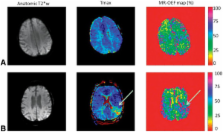
          Oxygenation-sensitive cardiovascular magnetic resonance (CMR) is a non-contrast technique that allows the non-invasive assessment of myocardial oxygenation. It capitalizes on the fact that deoxygenated hemoglobin in blood can act as an intrinsic contrast agent, changing proton signals in a fashion that can be imaged to reflect the level of blood oxygenation. Increases in O 2 saturation increase the BOLD imaging signal (T2 or T2*), whereas decreases diminish it. This review presents the basic concepts and limitations of the BOLD technique, and summarizes the preclinical and clinical studies in the assessment of myocardial oxygenation with a focus on recent advances. Finally, it provides future directions and a brief look at emerging techniques of this evolving CMR field.

          Related collections

          Most cited references37

          • Record: found
          • Abstract: found
          • Article: not found

          Intrinsic signal changes accompanying sensory stimulation: functional brain mapping with magnetic resonance imaging.

          We report that visual stimulation produces an easily detectable (5-20%) transient increase in the intensity of water proton magnetic resonance signals in human primary visual cortex in gradient echo images at 4-T magnetic-field strength. The observed changes predominantly occur in areas containing gray matter and can be used to produce high-spatial-resolution functional brain maps in humans. Reducing the image-acquisition echo time from 40 msec to 8 msec reduces the amplitude of the fractional signal change, suggesting that it is produced by a change in apparent transverse relaxation time T*2. The amplitude, sign, and echo-time dependence of these intrinsic signal changes are consistent with the idea that neural activation increases regional cerebral blood flow and concomitantly increases venous-blood oxygenation.
            Bookmark
            • Record: found
            • Abstract: not found
            • Article: not found

            The Magnetic Properties and Structure of Hemoglobin, Oxyhemoglobin and Carbonmonoxyhemoglobin.

              Bookmark
              • Record: found
              • Abstract: found
              • Article: not found

              In vivo measurement of T*2 and field inhomogeneity maps in the human heart at 1.5 T.

              Cardiac echo-planar imaging suffers invariably from regions of severe distortion and T*2 decay in the myocardium. The purpose of this work was to perform local measurements of T*2 and field inhomogeneities in the myocardium and to identify the sources of focal signal loss and distortion. Field inhomogeneity maps and T*2 were measured in five normal volunteers in short-axis slices spanning from base to apex. It was found that T*2 ranged from 26 ms (SD = 7 ms, n = 5) to 41 ms (SD = 11 ms, n = 5) over most of the heart, and peak-to-peak field inhomogeneity differences were 71 Hz (SD = 14 Hz, n = 5). In all hearts, regions of severe signal loss were consistently adjacent to the posterior vein of the left ventricle; T*2 in these regions was 12 ms (SD = 2 ms, n = 5), and the difference in resonance frequency with the surrounding myocardium was 70-100 Hz. These effects may be caused by increased magnetic susceptibility from deoxygenated blood in these veins.
                Bookmark

                Author and article information

                Journal
                J Cardiovasc Magn Reson
                J Cardiovasc Magn Reson
                Journal of Cardiovascular Magnetic Resonance
                BioMed Central
                1097-6647
                1532-429X
                2013
                24 May 2013
                : 15
                : 1
                : 43
                Affiliations
                [1 ]Montreal Heart Institute, Departments of Cardiology and Radiology, Université de Montréal, Montreal, QC, Canada
                [2 ]Departments of Cardiac Sciences and Radiology, University of Calgary, Calgary, Canada
                [3 ]Oxford Centre for Clinical Magnetic Resonance Research, Division of Cardiovascular Medicine, Radcliffe Department of Medicine, University of Oxford, John Radcliffe Hospital, Oxford, UK
                Article
                1532-429X-15-43
                10.1186/1532-429X-15-43
                3681671
                23706167
                c57d5de3-4d10-476b-8e6f-95ea4b8e8e50
                Copyright ©2013 Friedrich and Karamitsos; licensee BioMed Central Ltd.

                This is an Open Access article distributed under the terms of the Creative Commons Attribution License ( http://creativecommons.org/licenses/by/2.0), which permits unrestricted use, distribution, and reproduction in any medium, provided the original work is properly cited.

                History
                : 29 April 2013
                : 29 April 2013
                Categories
                Review

                Cardiovascular Medicine
                cardiovascular magnetic resonance,blood-oxygen level-dependent,microcirculation,ischemia,oxygenation

                Comments

                Comment on this article