125
views
0
recommends
+1 Recommend
0 collections
    12
    shares
      • Record: found
      • Abstract: found
      • Article: not found

      Extracellular histones are major mediators of death in sepsis

      research-article

      Read this article at

      ScienceOpenPublisherPMC
      Bookmark
          There is no author summary for this article yet. Authors can add summaries to their articles on ScienceOpen to make them more accessible to a non-specialist audience.

          Abstract

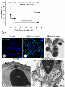
          Hyper–inflammatory responses can lead to a variety of diseases including sepsis 1. We now report that extracellular histones released in response to inflammatory challenge contribute to endothelial dysfunction, organ failure and death during sepsis. They can be targeted pharmacologically by antibody to histone or by activated protein C (APC). Antibody to histone reduced the mortality of mice in lipopolysaccharide (LPS), tumor necrosis factor (TNF) or cecal ligation and puncture models of sepsis. Extracellular histones are cytotoxic toward endothelium in vitro and are lethal in mice. In vivo, histone administration resulted in neutrophil margination, vacuolated endothelium, intra–alveolar hemorrhage and macro and microvascular thrombosis. Histone was detected in the circulation of baboons challenged with E. coli and the increase in histone levels accompanied the onset of renal dysfunction. APC cleaves histones and reduces their cytotoxicity. Co–infusion of APC with E. coli in baboons or histones in mice prevented lethality. Blockade of protein C activation exacerbated sublethal LPS challenge into lethality which was reversed by antibody to histone. We conclude that extracellular histones are potential molecular targets for therapeutics for sepsis and other inflammatory diseases.

          Related collections

          Most cited references19

          • Record: found
          • Abstract: found
          • Article: not found

          Platelet TLR4 activates neutrophil extracellular traps to ensnare bacteria in septic blood.

          It has been known for many years that neutrophils and platelets participate in the pathogenesis of severe sepsis, but the inter-relationship between these players is completely unknown. We report several cellular events that led to enhanced trapping of bacteria in blood vessels: platelet TLR4 detected TLR4 ligands in blood and induced platelet binding to adherent neutrophils. This led to robust neutrophil activation and formation of neutrophil extracellular traps (NETs). Plasma from severely septic humans also induced TLR4-dependent platelet-neutrophil interactions, leading to the production of NETs. The NETs retained their integrity under flow conditions and ensnared bacteria within the vasculature. The entire event occurred primarily in the liver sinusoids and pulmonary capillaries, where NETs have the greatest capacity for bacterial trapping. We propose that platelet TLR4 is a threshold switch for this new bacterial trapping mechanism in severe sepsis.
            Bookmark
            • Record: found
            • Abstract: found
            • Article: not found

            HMG-1 as a late mediator of endotoxin lethality in mice.

            Endotoxin, a constituent of Gram-negative bacteria, stimulates macrophages to release large quantities of tumor necrosis factor (TNF) and interleukin-1 (IL-1), which can precipitate tissue injury and lethal shock (endotoxemia). Antagonists of TNF and IL-1 have shown limited efficacy in clinical trials, possibly because these cytokines are early mediators in pathogenesis. Here a potential late mediator of lethality is identified and characterized in a mouse model. High mobility group-1 (HMG-1) protein was found to be released by cultured macrophages more than 8 hours after stimulation with endotoxin, TNF, or IL-1. Mice showed increased serum levels of HMG-1 from 8 to 32 hours after endotoxin exposure. Delayed administration of antibodies to HMG-1 attenuated endotoxin lethality in mice, and administration of HMG-1 itself was lethal. Septic patients who succumbed to infection had increased serum HMG-1 levels, suggesting that this protein warrants investigation as a therapeutic target.
              Bookmark
              • Record: found
              • Abstract: found
              • Article: not found

              Immunodesign of experimental sepsis by cecal ligation and puncture.

              Sepsis remains a prevalent clinical challenge and the underlying pathophysiology is still poorly understood. To investigate the complex molecular mechanisms of sepsis, various animal models have been developed, the most frequently used being the cecal ligation and puncture (CLP) model in rodents. In this model, sepsis originates from a polymicrobial infectious focus within the abdominal cavity, followed by bacterial translocation into the blood compartment, which then triggers a systemic inflammatory response. A requirement of this model is that it is performed with high consistency to obtain reproducible results. Evidence is now emerging that the accompanying inflammatory response varies with the severity grade of sepsis, which is highly dependent on the extent of cecal ligation. In this protocol, we define standardized procedures for inducing sepsis in mice and rats by applying defined severity grades of sepsis through modulation of the position of cecal ligation. The CLP procedure can be performed in as little as 10 min for each animal by an experienced user, with additional time required for subsequent postoperative care and data collection.
                Bookmark

                Author and article information

                Journal
                9502015
                8791
                Nat Med
                Nature medicine
                1078-8956
                1546-170X
                9 October 2009
                25 October 2009
                November 2009
                1 May 2010
                : 15
                : 11
                : 1318-1321
                Affiliations
                [1 ]Cardiovascular Biology Research Program, Oklahoma City, OK 73104
                [2 ]Howard Hughes Medical Institute, Oklahoma City, OK 73104
                [3 ]Immunology and Cancer Research Program, Oklahoma Medical Research Foundation, Temple University School of Medicine, Philadelphia, PA 19140, USA
                [4 ]Temple Autoimmunity Center, Department of Microbiology and Immunology, Temple University School of Medicine, Philadelphia, PA 19140, USA
                Author notes
                Correspondence and material request should be addressed to C. T. E. ( esmonc@ 123456omrf.org )
                Article
                nihpa149563
                10.1038/nm.2053
                2783754
                19855397
                c59638c8-a6e6-490a-8fd3-b92320853253

                Users may view, print, copy, download and text and data- mine the content in such documents, for the purposes of academic research, subject always to the full Conditions of use: http://www.nature.com/authors/editorial_policies/license.html#terms

                History
                Funding
                Funded by: National Institute of General Medical Sciences : NIGMS
                Award ID: R01 GM037704-19 ||GM
                Categories
                Article

                Medicine
                Medicine

                Comments

                Comment on this article