16
views
0
recommends
+1 Recommend
0 collections
    0
    shares
      • Record: found
      • Abstract: found
      • Article: not found

      Retroperitoneal Actinomycosis: A Rare Sequela of an Infected Obstructing Ureteral Stone

      case-report

      Read this article at

      ScienceOpenPublisherPMC
      Bookmark
          There is no author summary for this article yet. Authors can add summaries to their articles on ScienceOpen to make them more accessible to a non-specialist audience.

          Abstract

          Background: Actinomycosis is a condition in which Actinomyces, a normal component of the oral and gastrointenstial flora, becomes pathogenic in the setting of damaged tissue, leading to widespread tissue destruction across fascial planes. Prior literature describing this condition is rare, particularly cases involving the retroperitoneum. In this study, we report a case of retroperitoneal actinomycosis caused by an infected, obstructing ureteral stone.

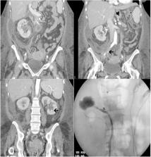
          Case Presentation: A 48-year-old woman with a history of substance abuse, malnutrition, and gastric bypass presented to the emergency room with a 3-week history of abdominal pain and fevers. Workup revealed a 9 mm obstructing right ureteral stone with associated perinephric fluid collection that was concerning for forniceal rupture. There was left hydronephrosis and a 3 mm lower pole renal calculus as well. The patient underwent emergent decompression where bilateral duplicated collecting systems were identified, requiring stenting of all four moieties to ensure maximal decompression in the setting of obstructive pyelonephritis. Urine cultures grew Escherichia coli and Candida. The patient continued to deteriorate despite culture appropriate antibiotic therapy; repeat scan revealed progression of her perinephric fluid collection into a loculated retroperitoneal abscess. A percutaneous drain was placed, and nearly half a liter of pus was evacuated. Fluid cultures grew Actinomyces, and she ultimately recovered after a prolonged course of antibiotics, including 1 month of intravenous therapy and an additional 6 months of oral treatment. All stones were ultimately removed via ureteroscopy.

          Conclusion: Actinomycosis is a rare invasive infection that is caused when the Actinomyces bacteria colonizes damaged tissue. We present the first reported case of urolithiasis inciting this process via tissue damage caused by obstruction and infection. Although rare, heightened suspicion is warranted among immunocompromised hosts who do not improve after decompression in such scenarios.

          Related collections

          Most cited references4

          • Record: found
          • Abstract: found
          • Article: found
          Is Open Access

          Actinomycosis: etiology, clinical features, diagnosis, treatment, and management

          Actinomycosis is a rare chronic disease caused by Actinomyces spp., anaerobic Gram-positive bacteria that normally colonize the human mouth and digestive and genital tracts. Physicians must be aware of typical clinical presentations (such as cervicofacial actinomycosis following dental focus of infection, pelvic actinomycosis in women with an intrauterine device, and pulmonary actinomycosis in smokers with poor dental hygiene), but also that actinomycosis may mimic the malignancy process in various anatomical sites. Bacterial cultures and pathology are the cornerstone of diagnosis, but particular conditions are required in order to get the correct diagnosis. Prolonged bacterial cultures in anaerobic conditions are necessary to identify the bacterium and typical microscopic findings include necrosis with yellowish sulfur granules and filamentous Gram-positive fungal-like pathogens. Patients with actinomycosis require prolonged (6- to 12-month) high doses (to facilitate the drug penetration in abscess and in infected tissues) of penicillin G or amoxicillin, but the duration of antimicrobial therapy could probably be shortened to 3 months in patients in whom optimal surgical resection of infected tissues has been performed. Preventive measures, such as reduction of alcohol abuse and improvement of dental hygiene, may limit occurrence of pulmonary, cervicofacial, and central nervous system actinomycosis. In women, intrauterine devices must be changed every 5 years in order to limit the occurrence of pelvic actinomycosis.
            Bookmark
            • Record: found
            • Abstract: found
            • Article: found
            Is Open Access

            Clinical features of actinomycosis

            Abstract Actinomycosis is a rare heterogeneous anaerobic infection with misleading clinical presentations that delay diagnosis. A significant number of misdiagnosed cases have been reported in specific localizations, but studies including various forms of actinomycosis have rarely been published. We performed a multicenter retrospective chart review of laboratory-confirmed actinomycosis cases from January 2000 until January 2014. We described clinical characteristics, diagnostic procedures, differential diagnosis, and management of actinomycosis of clinical significance. Twenty-eight patients were included from 6 hospitals in France. Disease was diagnosed predominately in the abdomen/pelvis (n = 9), orocervicofacial (n = 5), cardiothoracic (n = 5), skeletal (n = 3), hematogenous (n = 3), soft tissue (n = 2), and intracranially (n = 1). Four patients (14%) were immunocompromised. In most cases (92 %), the diagnosis of actinomycosis was not suspected on admission, as clinical features were not specific. Diagnosis was obtained from either microbiology (50%, n = 14) or histopathology (42%, n = 12), or from both methods (7%, n = 2). Surgical biopsy was needed for definite diagnosis in 71% of cases (n = 20). Coinfection was found in 13 patients (46%), among which 3 patients were diagnosed from histologic criteria only. Two-thirds of patients were treated with amoxicillin. Median duration of antibiotics was 120 days (interquartile range 60–180), whereas the median follow-up time was 12 months (interquartile range 5.25–18). Two patients died. This study highlights the distinct and miscellaneous patterns of actinomycosis to prompt accurate diagnosis and earlier treatments, thus improving the outcome. Surgical biopsy should be performed when possible while raising histologist's and microbiologist's awareness of possible actinomycosis to enhance the chance of diagnosis and use specific molecular methods.
              Bookmark
              • Record: found
              • Abstract: found
              • Article: not found

              Recognition of renal actinomycosis: nephrectomy can be avoided. Report of a case.

              Nephrectomy is performed for the diagnosis and treatment of renal actinomycosis. A cachectic 62-year-old man with a left renal mass underwent exploration and needle biopsy of the mass. No malignant disease was found. When his condition further deteriorated, the kidney was re-explored. Numerous biopsies intraoperatively finally revealed actinomycosis, so the operation was terminated with preservation of the kidney. Long-term treatment with antibiotics was begun with immediate, marked improvement in the patient's condition. At 1 year the renal mass had almost resolved. This is the first reported case of the diagnosis and treatment of renal actinomycosis without nephrectomy.
                Bookmark

                Author and article information

                Journal
                J Endourol Case Rep
                J Endourol Case Rep
                cren
                Journal of Endourology Case Reports
                Mary Ann Liebert, Inc. (140 Huguenot Street, 3rd FloorNew Rochelle, NY 10801USA )
                2379-9889
                01 July 2017
                2017
                01 July 2017
                : 3
                : 1
                : 93-96
                Affiliations
                [ 1 ]Department of Urology, University of Minnesota Medical School , Minneapolis, Minnesota.
                [ 2 ]Division of Infectious Diseases and International Medicine, Department of Medicine, University of Minnesota Medical School , Minneapolis, Minnesota.
                Author notes
                Address correspondence to: Michael S. Borofsky, MD, Department of Urology, University of Minnesota Medical School 420 Delaware Street SE, Box 394 Mayo, Minneapolis, MN 55455, E-mail: mborofsk@ 123456umn.edu
                Article
                10.1089/cren.2017.0048
                10.1089/cren.2017.0048
                5587902
                28894842
                c5c8f1c1-2cb4-4791-9c92-ea35f0973ed9
                © Elizabeth Bearrick et al. 2017; Published by Mary Ann Liebert, Inc.

                This is an Open Access article distributed under the terms of the Creative Commons Attribution License, which permits unrestricted use, distribution, and reproduction in any medium, provided the original work is properly cited.

                History
                Page count
                Figures: 2, References: 4, Pages: 4
                Categories
                Case Report

                ureteral stones,infection/inflammation,urolithiasis,ureteroscopy,actinomycosis,pyelonephritis,retroperitoneum

                Comments

                Comment on this article