2
views
0
recommends
+1 Recommend
0 collections
    0
    shares
      • Record: found
      • Abstract: found
      • Article: found
      Is Open Access

      Clinical and Genetic Characteristics of Chinese Patients with Occult Macular Dystrophy

      research-article

      Read this article at

      Bookmark
          There is no author summary for this article yet. Authors can add summaries to their articles on ScienceOpen to make them more accessible to a non-specialist audience.

          Abstract

          Purpose

          To investigate the clinical and genetic characteristics of occult macular dystrophy (OMD) based on a Chinese patient cohort.

          Methods

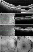
          Fifteen Chinese OMD patients from nine unrelated families underwent genetic testing, and all of them harbored a pathogenic RP1L1 variant. Comprehensive ophthalmic examinations were performed in nine probands, including spectral-domain optical coherence tomography (SD-OCT), near-infrared reflectance (NIR), fundus autofluorescence (AF), and multifocal electroretinography.

          Results

          The RP1L1 variants p.R45W and p.S1199C were identified in 13 patients and two patients, respectively, and one was a de novo mutation. Among the nine probands, the median ages at onset and examination were 25.0 years (range, 6–51 years) and 27.0 years (range, 14–55 years), respectively. The median decimal visual acuity was 0.20 (range, 0.04–0.5). Foveal photoreceptor thickness and visual acuity showed a significant correlation ( r = 0.591; P = 0.01). All eyes presented with an absent interdigitation zone and blurred ellipsoid zone of photoreceptors when examined by SD-OCT. In addition, central round lesions with low NIR reflectance were observed in 66.7% (12/18) of eyes by NIR reflectance imaging, corresponding to the regions with abnormal photoreceptor microstructures observed by SD-OCT. Of the 18 eyes, only four eyes showed ring-like faint hyperfluorescence around the macula by AF.

          Conclusions

          To the best of our knowledge, this is the largest study in a cohort of Chinese OMD patients with RP1L1 mutations. Our findings revealed that the two recurrent RP1L1 variants are related to OMD in the Chinese population. Furthermore, multimodal imaging combined with genetic testing is valuable for diagnosing and monitoring OMD progression.

          Related collections

          Most cited references20

          • Record: found
          • Abstract: found
          • Article: found

          RP1L1 variants are associated with a spectrum of inherited retinal diseases including retinitis pigmentosa and occult macular dystrophy.

          In one consanguineous family with retinitis pigmentosa (RP), a condition characterized by progressive visual loss due to retinal degeneration, homozygosity mapping, and candidate gene sequencing suggested a novel locus. Exome sequencing identified a homozygous frameshifting mutation, c.601delG, p.Lys203Argfs*28, in RP1L1 encoding RP 1-like1, a photoreceptor-specific protein. A screen of a further 285 unrelated individuals with autosomal recessive RP identified an additional proband, homozygous for a missense variant, c.1637G>C, p.Ser546Thr, in RP1L1. A distinct retinal disorder, occult macular dystrophy (OCMD) solely affects the central retinal cone photoreceptors and has previously been reported to be associated with variants in the same gene. The association between mutations in RP1L1 and the disorder OCMD was explored by screening a cohort of 28 unrelated individuals with the condition; 10 were found to harbor rare (minor allele frequency ≤0.5% in the 1,000 genomes dataset) heterozygous RP1L1 missense variants. Analysis of family members revealed many unaffected relatives harboring the same variant. Linkage analysis excluded the possibility of a recessive mode of inheritance, and sequencing of RP1, a photoreceptor protein that interacts with RP1L1, excluded a digenic mechanism involving this gene. These findings imply an important and diverse role for RP1L1 in human retinal physiology and disease. © 2012 Wiley Periodicals, Inc.
            Bookmark
            • Record: found
            • Abstract: found
            • Article: not found

            Dominant mutations in RP1L1 are responsible for occult macular dystrophy.

            Occult macular dystrophy (OMD) is an inherited macular dystrophy characterized by progressive loss of macular function but normal ophthalmoscopic appearance. Typical OMD is characterized by a central cone dysfunction leading to a loss of vision despite normal ophthalmoscopic appearance, normal fluorescein angiography, and normal full-field electroretinogram (ERGs), but the amplitudes of the focal macular ERGs and multifocal ERGs are significantly reduced at the central retina. Linkage analysis of two OMD families was performed by the SNP High Throughput Linkage analysis system (SNP HiTLink), localizing the disease locus to chromosome 8p22-p23. Among the 128 genes in the linkage region, 22 genes were expressed in the retina, and four candidate genes were selected. No mutations were found in the first three candidate genes, methionine sulfoxide reductase A (MSRA), GATA binding 4 (GATA4), and pericentriolar material 1 (PCM1). However, amino acid substitution of p.Arg45Trp in retinitis pigmentosa 1-like 1 (RP1L1) was found in three OMD families and p.Trp960Arg in a remaining OMD family. These two mutations were detected in all affected individuals but in none of the 876 controls. Immunohistochemistry of RP1L1 in the retina section of cynomolgus monkey revealed expression in the rod and cone photoreceptor, supporting a role of RP1L1 in the photoreceptors that, when disrupted by mutation, leads to OMD. Identification of RP1L1 mutations as causative for OMD has potentially broader implications for understanding the differential cone photoreceptor functions in the fovea and the peripheral retina. 2010 The American Society of Human Genetics. Published by Elsevier Inc. All rights reserved.
              Bookmark
              • Record: found
              • Abstract: found
              • Article: not found

              Essential and synergistic roles of RP1 and RP1L1 in rod photoreceptor axoneme and retinitis pigmentosa.

              Retinitis pigmentosa 1 (RP1) is a common inherited retinopathy with variable onset and severity. The RP1 gene encodes a photoreceptor-specific, microtubule-associated ciliary protein containing the doublecortin (DCX) domain. Here we show that another photoreceptor-specific Rp1-like protein (Rp1L1) in mice is also localized to the axoneme of outer segments (OSs) and connecting cilia in rod photoreceptors, overlapping with Rp1. Rp1L1-/- mice display scattered OS disorganization, reduced electroretinogram amplitudes, and progressive photoreceptor degeneration, less severe and slower than in Rp1-/- mice. In single rods of Rp1L1-/-, photosensitivity is reduced, similar to that of Rp1-/-. While individual heterozygotes are normal, double heterozygotes of Rp1 and Rp1L1 exhibit abnormal OS morphology and reduced single rod photosensitivity and dark currents. The electroretinogram amplitudes of double heterozygotes are more reduced than those of individual heterozygotes combined. In support, Rp1L1 interacts with Rp1 in transfected cells and in retina pull-down experiments. Interestingly, phototransduction kinetics are normal in single rods and whole retinas of individual or double Rp1 and Rp1L1 mutant mice. Together, Rp1 and Rp1L1 play essential and synergistic roles in affecting photosensitivity and OS morphogenesis of rod photoreceptors. Our findings suggest that mutations in RP1L1 could underlie retinopathy or modify RP1 disease expression in humans.
                Bookmark

                Author and article information

                Journal
                Invest Ophthalmol Vis Sci
                Invest. Ophthalmol. Vis. Sci
                iovs
                IOVS
                Investigative Ophthalmology & Visual Science
                The Association for Research in Vision and Ophthalmology
                0146-0404
                1552-5783
                16 March 2020
                March 2020
                : 61
                : 3
                : 10
                Affiliations
                [1 ] Eye and ENT Hospital, Shanghai Medical College, Fudan University, Shanghai, China
                [2 ] Shanghai Key Laboratory of Visual Impairment and Restoration, Science and Technology Commission of Shanghai Municipality , Shanghai, China
                [3 ] Key Laboratory of Myopia, Ministry of Health , Shanghai, China
                [4 ] BGI-Shenzhen , Shenzhen, China
                [5 ] Sonoma Academy , California, USA
                Author notes
                Correspondence: Ji-Hong Wu, Eye and ENT Hospital of Fudan University, 83 Fengyang Road, Shanghai, 200031, China; jihongwu@ 123456fudan.edu.cn .
                Guo-Hong Tian, Eye, Ear, Nose and Throat Hospital of Fudan University, 83 Fengyang Road, Shanghai 200031, China; valentian99@ 123456hotmail.com .

                D-DW and F-JG contributed equally to this work.

                Article
                IOVS-19-28434
                10.1167/iovs.61.3.10
                7401461
                32176261
                c7551767-84a1-484d-b38e-711ea55ee121
                Copyright 2020 The Authors

                This work is licensed under a Creative Commons Attribution-NonCommercial-NoDerivatives 4.0 International License.

                History
                : 19 December 2019
                : 27 September 2019
                Page count
                Pages: 8
                Categories
                Retina
                Retina

                occult macular dystrophy,rp1l1,multimodal imaging,next-generation sequencing,early diagnostics

                Comments

                Comment on this article