79
views
0
recommends
+1 Recommend
0 collections
    0
    shares
      • Record: found
      • Abstract: found
      • Article: found
      Is Open Access

      PDL1 Regulation by p53 via miR-34

      research-article

      Read this article at

      Bookmark
          There is no author summary for this article yet. Authors can add summaries to their articles on ScienceOpen to make them more accessible to a non-specialist audience.

          Abstract

          Background:

          Although clinical studies have shown promise for targeting PD1/PDL1 signaling in non–small cell lung cancer (NSCLC), the regulation of PDL1 expression is poorly understood. Here, we show that PDL1 is regulated by p53 via miR-34.

          Methods:

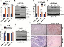
          p53 wild-type and p53-deficient cell lines (p53 –/– and p53 +/+ HCT116, p53-inducible H1299, and p53-knockdown H460) were used to determine if p53 regulates PDL1 via miR-34. PDL1 and miR-34a expression were analyzed in samples from patients with NSCLC and mutated p53 vs wild-type p53 tumors from The Cancer Genome Atlas for Lung Adenocarcinoma (TCGA LUAD). We confirmed that PDL1 is a direct target of miR-34 with western blotting and luciferase assays and used a p53 R172HΔg/+K-ras LA1/+ syngeneic mouse model (n = 12) to deliver miR-34a–loaded liposomes (MRX34) plus radiotherapy (XRT) and assessed PDL1 expression and tumor-infiltrating lymphocytes (TILs). A two-sided t test was applied to compare the mean between different treatments.

          Results:

          We found that p53 regulates PDL1 via miR-34, which directly binds to the PDL1 3’ untranslated region in models of NSCLC (fold-change luciferase activity to control group, mean for miR-34a = 0.50, SD = 0.2, P < .001; mean for miR-34b = 0.52, SD = 0.2, P = .006; and mean for miR-34c = 0.59, SD = 0.14, and P = .006). Therapeutic delivery of MRX34, currently the subject of a phase I clinical trial, promoted TILs (mean of CD8 expression percentage of control group = 22.5%, SD = 1.9%; mean of CD8 expression percentage of MRX34 = 30.1%, SD = 3.7%, P = .016, n = 4) and reduced CD8 +PD1 + cells in vivo (mean of CD8/PD1 expression percentage of control group = 40.2%, SD = 6.2%; mean of CD8/PD1 expression percentage of MRX34 = 20.3%, SD = 5.1%, P = .001, n = 4). Further, MRX34 plus XRT increased CD8 + cell numbers more than either therapy alone (mean of CD8 expression percentage of MRX34 plus XRT to control group = 44.2%, SD = 8.7%, P = .004, n = 4). Finally, miR-34a delivery reduced the numbers of radiation-induced macrophages (mean of F4-80 expression percentage of control group = 52.4%, SD = 1.7%; mean of F4-80 expression percentage of MRX34 = 40.1%, SD = 3.5%, P = .008, n = 4) and T-regulatory cells.

          Conclusions:

          We identified a novel mechanism by which tumor immune evasion is regulated by p53/miR-34/PDL1 axis. Our results suggest that delivery of miRNAs with standard therapies, such as XRT, may represent a novel therapeutic approach for lung cancer.

          Related collections

          Most cited references35

          • Record: found
          • Abstract: found
          • Article: not found

          The blockade of immune checkpoints in cancer immunotherapy.

          Among the most promising approaches to activating therapeutic antitumour immunity is the blockade of immune checkpoints. Immune checkpoints refer to a plethora of inhibitory pathways hardwired into the immune system that are crucial for maintaining self-tolerance and modulating the duration and amplitude of physiological immune responses in peripheral tissues in order to minimize collateral tissue damage. It is now clear that tumours co-opt certain immune-checkpoint pathways as a major mechanism of immune resistance, particularly against T cells that are specific for tumour antigens. Because many of the immune checkpoints are initiated by ligand-receptor interactions, they can be readily blocked by antibodies or modulated by recombinant forms of ligands or receptors. Cytotoxic T-lymphocyte-associated antigen 4 (CTLA4) antibodies were the first of this class of immunotherapeutics to achieve US Food and Drug Administration (FDA) approval. Preliminary clinical findings with blockers of additional immune-checkpoint proteins, such as programmed cell death protein 1 (PD1), indicate broad and diverse opportunities to enhance antitumour immunity with the potential to produce durable clinical responses.
            Bookmark
            • Record: found
            • Abstract: found
            • Article: not found

            Involvement of PD-L1 on tumor cells in the escape from host immune system and tumor immunotherapy by PD-L1 blockade.

            PD-1 is a receptor of the Ig superfamily that negatively regulates T cell antigen receptor signaling by interacting with the specific ligands (PD-L) and is suggested to play a role in the maintenance of self-tolerance. In the present study, we examined possible roles of the PD-1/PD-L system in tumor immunity. Transgenic expression of PD-L1, one of the PD-L, in P815 tumor cells rendered them less susceptible to the specific T cell antigen receptor-mediated lysis by cytotoxic T cells in vitro, and markedly enhanced their tumorigenesis and invasiveness in vivo in the syngeneic hosts as compared with the parental tumor cells that lacked endogenous PD-L. Both effects could be reversed by anti-PD-L1 Ab. Survey of murine tumor lines revealed that all of the myeloma cell lines examined naturally expressed PD-L1. Growth of the myeloma cells in normal syngeneic mice was inhibited significantly albeit transiently by the administration of anti-PD-L1 Ab in vivo and was suppressed completely in the syngeneic PD-1-deficient mice. These results suggest that the expression of PD-L1 can serve as a potent mechanism for potentially immunogenic tumors to escape from host immune responses and that blockade of interaction between PD-1 and PD-L may provide a promising strategy for specific tumor immunotherapy.
              Bookmark
              • Record: found
              • Abstract: found
              • Article: not found

              Senescence and tumour clearance is triggered by p53 restoration in murine liver carcinomas.

              Although cancer arises from a combination of mutations in oncogenes and tumour suppressor genes, the extent to which tumour suppressor gene loss is required for maintaining established tumours is poorly understood. p53 is an important tumour suppressor that acts to restrict proliferation in response to DNA damage or deregulation of mitogenic oncogenes, by leading to the induction of various cell cycle checkpoints, apoptosis or cellular senescence. Consequently, p53 mutations increase cell proliferation and survival, and in some settings promote genomic instability and resistance to certain chemotherapies. To determine the consequences of reactivating the p53 pathway in tumours, we used RNA interference (RNAi) to conditionally regulate endogenous p53 expression in a mosaic mouse model of liver carcinoma. We show that even brief reactivation of endogenous p53 in p53-deficient tumours can produce complete tumour regressions. The primary response to p53 was not apoptosis, but instead involved the induction of a cellular senescence program that was associated with differentiation and the upregulation of inflammatory cytokines. This program, although producing only cell cycle arrest in vitro, also triggered an innate immune response that targeted the tumour cells in vivo, thereby contributing to tumour clearance. Our study indicates that p53 loss can be required for the maintenance of aggressive carcinomas, and illustrates how the cellular senescence program can act together with the innate immune system to potently limit tumour growth.
                Bookmark

                Author and article information

                Journal
                J Natl Cancer Inst
                J. Natl. Cancer Inst
                jnci
                jnci
                JNCI Journal of the National Cancer Institute
                Oxford University Press (US )
                0027-8874
                1460-2105
                January 2016
                17 November 2015
                17 November 2015
                : 108
                : 1
                : djv303
                Affiliations
                Affiliations of authors: Departments of Experimental Radiation Oncology (MAC, DV, XW, YY), Experimental Therapeutics (CI, GAC), and Radiation Oncology (RK, DRG, SK, JWW), The University of Texas MD Anderson Cancer Center , Houston, TX; Mirna Therapeutics, Inc. , Austin, TX (HJP, KK, DM, AGB); Ohio State University , Columbus, OH (LA, DPC, KS); Texas Veterinary Pathology Associates (Houston) , Houston, TX (DKG).
                Author notes
                Correspondence to: James W. Welsh, MD, Department of Radiation Oncology, Unit 97, The University of Texas MD Anderson Cancer Center, 1515 Holcombe Blvd, Houston, TX 77030 (e-mail: jwelsh@ 123456mdanderson.org ).
                Article
                10.1093/jnci/djv303
                4862407
                26577528
                c78b1b20-0341-4942-a5af-420c4cef8ca9
                © The Author 2015. Published by Oxford University Press.

                This is an Open Access article distributed under the terms of the Creative Commons Attribution License ( http://creativecommons.org/licenses/by/3.0/), which permits unrestricted reuse, distribution, and reproduction in any medium, provided the original work is properly cited.

                History
                : 16 December 2014
                : 29 April 2015
                : 25 September 2015
                Page count
                Pages: 9
                Funding
                Funded by: Doctors Cancer Foundation http://dx.doi.org/10.13039/100005689
                Funded by: Lung Cancer Research Foundation http://dx.doi.org/10.13039/100007038
                Award ID: Grant CA016672
                Categories
                Article

                Oncology & Radiotherapy
                Oncology & Radiotherapy

                Comments

                Comment on this article