34
views
0
recommends
+1 Recommend
0 collections
    0
    shares
      • Record: found
      • Abstract: found
      • Article: not found

      Kinesin-1 structural organization and conformational changes revealed by FRET stoichiometry in live cells

      research-article

      Read this article at

      ScienceOpenPublisherPMC
      Bookmark
          There is no author summary for this article yet. Authors can add summaries to their articles on ScienceOpen to make them more accessible to a non-specialist audience.

          Abstract

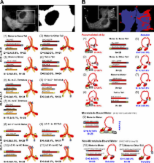
          Kinesin motor proteins drive the transport of cellular cargoes along microtubule tracks. How motor protein activity is controlled in cells is unresolved, but it is likely coupled to changes in protein conformation and cargo association. By applying the quantitative method fluorescence resonance energy transfer (FRET) stoichiometry to fluorescent protein (FP)–labeled kinesin heavy chain (KHC) and kinesin light chain (KLC) subunits in live cells, we studied the overall structural organization and conformation of Kinesin-1 in the active and inactive states. Inactive Kinesin-1 molecules are folded and autoinhibited such that the KHC tail blocks the initial interaction of the KHC motor with the microtubule. In addition, in the inactive state, the KHC motor domains are pushed apart by the KLC subunit. Thus, FRET stoichiometry reveals conformational changes of a protein complex in live cells. For Kinesin-1, activation requires a global conformational change that separates the KHC motor and tail domains and a local conformational change that moves the KHC motor domains closer together.

          Related collections

          Most cited references41

          • Record: found
          • Abstract: found
          • Article: not found

          RADIOAUTOGRAPHIC STUDIES OF CHOLINE INCORPORATION INTO PERIPHERAL NERVE MYELIN

          This radioautographic study was designed to localize the cytological sites involved in the incorporation of a lipid precursor into the myelin and the myelin-related cell of the peripheral nervous system. Both myelinating and fully myelinated cultures of rat dorsal root ganglia were exposed to a 30-min pulse of tritiated choline and either fixed immediately or allowed 6 or 48 hr of chase incubation before fixation. After Epon embedding, light and electron microscopic radioautograms were prepared with Ilford L-4 emulsion. Analysis of the pattern of choline incorporation into myelinating cultures indicated that radioactivity appeared all along the length of the internode, without there being a preferential site of initial incorporation. Light microscopic radioautograms of cultures at varying states of maturity were compared in order to determine the relative degree of myelin labeling. This analysis indicated that the myelin-Schwann cell unit in the fully myelinated cultures incorporated choline as actively as did this unit in the myelinating cultures. Because of technical difficulties, it was not possible to determine the precise localization of the incorporated radioactivity within the compact myelin. These data are related to recent biochemical studies indicating that the mature myelin of the central nervous system does incorporate a significant amount of lipid precursor under the appropriate experimental conditions. These observations support the concept that a significant amount of myelin-related metabolic activity occurs in mature tissue; this activity is considered part of an essential and continuous process of myelin maintenance and repair.
            Bookmark
            • Record: found
            • Abstract: found
            • Article: not found

            The green fluorescent protein.

            R Tsien (1998)
            In just three years, the green fluorescent protein (GFP) from the jellyfish Aequorea victoria has vaulted from obscurity to become one of the most widely studied and exploited proteins in biochemistry and cell biology. Its amazing ability to generate a highly visible, efficiently emitting internal fluorophore is both intrinsically fascinating and tremendously valuable. High-resolution crystal structures of GFP offer unprecedented opportunities to understand and manipulate the relation between protein structure and spectroscopic function. GFP has become well established as a marker of gene expression and protein targeting in intact cells and organisms. Mutagenesis and engineering of GFP into chimeric proteins are opening new vistas in physiological indicators, biosensors, and photochemical memories.
              Bookmark
              • Record: found
              • Abstract: found
              • Article: not found

              Molecular motors.

              Life implies movement. Most forms of movement in the living world are powered by tiny protein machines known as molecular motors. Among the best known are motors that use sophisticated intramolecular amplification mechanisms to take nanometre steps along protein tracks in the cytoplasm. These motors transport a wide variety of cargo, power cell locomotion, drive cell division and, when combined in large ensembles, allow organisms to move. Motor defects can lead to severe diseases or may even be lethal. Basic principles of motor design and mechanism have now been derived, and an understanding of their complex cellular roles is emerging.
                Bookmark

                Author and article information

                Journal
                J Cell Biol
                jcb
                The Journal of Cell Biology
                The Rockefeller University Press
                0021-9525
                1540-8140
                1 January 2007
                : 176
                : 1
                : 51-63
                Affiliations
                [1 ]Biophysics Research Division, [2 ]Department of Cell and Developmental Biology, and [3 ]Department of Microbiology and Immunology, University of Michigan, Ann Arbor, MI 48109
                Author notes

                Correspondence to Kristen J. Verhey: kjverhey@ 123456umich.edu

                Article
                200605097
                10.1083/jcb.200605097
                2063625
                17200416
                c7f08d6d-6496-414c-9220-f301cca61177
                Copyright © 2007, The Rockefeller University Press
                History
                : 16 May 2006
                : 1 December 2006
                Categories
                Research Articles
                Article

                Cell biology
                Cell biology

                Comments

                Comment on this article