13
views
0
recommends
+1 Recommend
0 collections
    0
    shares
      • Record: found
      • Abstract: found
      • Article: not found

      Nephrocalcinosis is a risk factor for kidney failure in primary hyperoxaluria

      research-article

      Read this article at

      ScienceOpenPublisherPMC
      Bookmark
          There is no author summary for this article yet. Authors can add summaries to their articles on ScienceOpen to make them more accessible to a non-specialist audience.

          Abstract

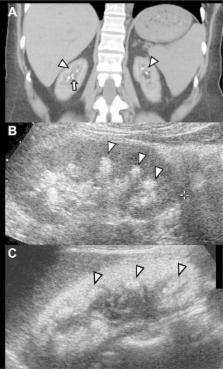
          Stone formation and nephrocalcinosis are both very common features of primary hyperoxaluria, yet the extent of each disease varies markedly between patients. Here we studied whether kidney damage from nephrocalcinosis and/or stone related events contributed to end stage kidney disease (ESKD). Clinical information was analyzed from 348 patients enrolled in the Rare Kidney Stone Consortium Primary Hyperoxaluria registry and included demographic, laboratory and imaging features. Among all patients there were 277 with type 1, 37 with type 2, and 34 with type 3 primary hyperoxaluria. Overall, 58% passed a stone (mean 0.3/year) and one or more urologic procedures were required by 70% of patients (mean 0.15/year). Nephrocalcinosis was found in 34% of patients, including 41% with type 1 primary hyperoxaluria. High urine oxalate was associated with increased risk for both nephrocalcinosis and stone number, while low urine citrate was a risk factor for stone events and stone number. After adjustment for the type of primary hyperoxaluria, diagnosis by family screening and age at first image, the overall adjusted hazard ratio for ESKD among those with a history of nephrocalcinosis was 1.7 [95% CI 1.0–3.0], while the risk was 4.0 [1.9–8.5] for new onset nephrocalcinosis during follow-up. In contrast, the number of stones and stone events were not significantly associated with ESKD risk. Thus, nephrolithiasis and nephrocalcinosis appear to be pathophysiologically distinct entities. The presence of nephrocalcinosis implies increased risk for ESKD.

          Related collections

          Most cited references18

          • Record: found
          • Abstract: found
          • Article: not found

          Hereditary causes of kidney stones and chronic kidney disease.

          Adenine phosphoribosyltransferase (APRT) deficiency, cystinuria, Dent disease, familial hypomagnesemia with hypercalciuria and nephrocalcinosis (FHHNC), and primary hyperoxaluria (PH) are rare but important causes of severe kidney stone disease and/or chronic kidney disease in children. Recurrent kidney stone disease and nephrocalcinosis, particularly in pre-pubertal children, should alert the physician to the possibility of an inborn error of metabolism as the underlying cause. Unfortunately, the lack of recognition and knowledge of the five disorders has frequently resulted in an unacceptable delay in diagnosis and treatment, sometimes with grave consequences. A high index of suspicion coupled with early diagnosis may reduce or even prevent the serious long-term complications of these diseases. In this paper, we review the epidemiology, clinical features, diagnosis, treatment, and outcome of patients with APRT deficiency, cystinuria, Dent disease, FHHNC, and PH, with an emphasis on childhood manifestations.
            Bookmark
            • Record: found
            • Abstract: found
            • Article: not found

            Calcium oxalate crystal adherence to hyaluronan-, osteopontin-, and CD44-expressing injured/regenerating tubular epithelial cells in rat kidneys.

            Retention of crystals in the kidney is an essential early step in renal stone formation. Studies with renal tubular cells in culture indicate that hyaluronan (HA) and osteopontin (OPN) and their mutual cell surface receptor CD44 play an important role in calcium oxalate (CaOx) crystal binding during wound healing. This concept was investigated in vivo by treating rats for 1, 4, and 8 d with ethylene glycol (0.5 and 0.75%) in their drinking water to induce renal tubular cell damage and CaOx crystalluria. Tubular injury was morphologically scored on periodic acid-Schiff-stained renal tissue sections and tissue repair assessed by immunohistochemical staining for proliferating cell nuclear antigen. CaOx crystals were visualized in periodic acid-Schiff-stained sections by polarized light microscopy, and renal calcium deposits were quantified with von Kossa staining. HA was visualized with HA-binding protein and OPN and CD44 immunohistochemically with specific antibodies and quantified with an image analyzer system. Already after 1 d of treatment, both concentrations of ethylene glycol induced hyperoxaluria and CaOx crystalluria. At this point, there was neither tubular injury nor crystal retention in the kidney, and expression of HA, OPN, and CD44 was comparable to untreated controls. After 4 and 8 d of ethylene glycol, however, intratubular crystals were found adhered to injured/regenerating (proliferating cell nuclear antigen positive) tubular epithelial cells, expressing HA, OPN, and CD44 at their luminal membrane. In conclusion, the expression of HA, OPN, and CD44 by injured/regenerating tubular cells seems to play a role in retention of crystals in the rat kidney.
              Bookmark
              • Record: found
              • Abstract: found
              • Article: not found

              EQUIL2: a BASIC computer program for the calculation of urinary saturation.

              A BASIC computer program for the calculation of urinary supersaturation with respect to the common kidney stone components is described. In vitro and in vivo tests show that the program described accurately calculates supersaturation. The application of this computer program to urolithiasis research is discussed.
                Bookmark

                Author and article information

                Journal
                0323470
                5428
                Kidney Int
                Kidney Int.
                Kidney international
                0085-2538
                1523-1755
                12 August 2014
                17 September 2014
                March 2015
                01 September 2015
                : 87
                : 3
                : 623-631
                Affiliations
                [1 ]Division of Nephrology and Hypertension, Mayo Clinic, Rochester, MN
                [2 ]Division of Biostatistics, Mayo Clinic, Rochester, MN
                [3 ]Department of Radiology, Mayo Clinic, Rochester, MN
                [4 ]Department of Laboratory Medicine and Pathology, Mayo Clinic, Rochester, MN
                [5 ]Division of Nephrology, Shanghai Changzheng Hospital, Second Military Medical University, Shanghai, China
                Author notes
                Corresponding author: John C. Lieske, MD, Mayo Clinic Division of Nephrology and Hypertension, 200 First St SW, Rochester, MN 55905, tel 507-266-7960;, fax 507-284-9315, Lieske.John@ 123456mayo.edu
                Article
                NIHMS619470
                10.1038/ki.2014.298
                4344931
                25229337
                c84234c7-1971-47bf-9097-362baf0184eb
                History
                Categories
                Article

                Nephrology
                end stage kidney disease,nephrocalcinosis,nephrolithiasis,oxalate,primary hyperoxaluria
                Nephrology
                end stage kidney disease, nephrocalcinosis, nephrolithiasis, oxalate, primary hyperoxaluria

                Comments

                Comment on this article