16
views
0
recommends
+1 Recommend
0 collections
    0
    shares
      • Record: found
      • Abstract: found
      • Article: found
      Is Open Access

      Ultra wide field imaging of coats like response in Leber’s congenital amaurosis

      brief-report
      * , , ,
      Saudi Journal of Ophthalmology
      Elsevier

      Read this article at

      Bookmark
          There is no author summary for this article yet. Authors can add summaries to their articles on ScienceOpen to make them more accessible to a non-specialist audience.

          Abstract

          A 12-year-old boy presented with poor vision and abnormal eye movements in his both eyes since birth. There were no systemic complaints. The visual acuity was light perception only in both eyes. The cycloplegic refraction revealed a hypermetropia of 6 and 7 diopters in right and left eye respectively, but the correction did not improve his visual acuity. The child had pendular nystagmus; enophthalmos and oculo-digital sign were present. The pupil reacted sluggishly to light in both the eyes. A dilated fundus examination revealed bilateral optic disc pallor, pigment spicules all over the fundus 
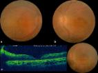
and vascular attenuation. In addition the right eye (Fig. 1) showed well-defined excavated area of chorio-retinal atrophy in macula and sclerosed vessels with scattered retinal hemorrhages in the temporal fundus. The left eye showed thick glial tissue with exudation in macula (Fig. 2). An area of retinal vascular telangiectasias and subretinal exudation was noted inferiorly and temporally in left fundus. Electroretinography (ERG) showed extinguished scotopic and photopic responses. A diagnosis of Leber’s Congenital Amaurosis (LCA) with “Coats like” response was made. Patient was advised for regular follow-up. Figure 1 Ultra wide field pseudo colour image of the right eye showing disc pallor, wide spread pigmentary changes, macular excavation and sclerosed vessels in temporal periphery. Figure 2 Pseudo colour image of left eye showing disc pallor, pigmentary changes and fibro-glial nodule at macula. In addition subretinal and retinal exudations along with telangiectasia are seen in temporal and infero-temporal area. Discussion Leber’s congenital amaurosis is a severe hereditary retinal dystrophy that leads to blindness manifesting in infancy. It is believed to be a cause of childhood blindness in as many as 20% cases in blind institutes and accounts for 5% of all retinal dystrophies. 1 It is mostly inherited as an autosomal recessive trait and to date around 19 genes have been implicated in the causation. Still, only around 70% or fewer cases are documented to have the detailed genetic defects. 2 The fundus appearance in LCA can be extremely variable. Most cases initially present with a normal fundus appearance or with mild granularity and vascular attenuation, though in later life a number of changes have been described. Typical fundal changes include macular coloboma, typical retinitis pigmentosa, retinitis punctata albescens, optic atrophy, salt and pepper retinopathy, peripheral nummular pigmentation and vascular complications, such as optic disc oedema, retinal vasculitis, secondary angiomatosis and astrocytoma. “Coats’ like response” is a clinical entity different from typical Coats disease and can be seen in a variety of clinical scenarios. It is typically described as a vascular alterations (telangiectasia and aneurysmal dilatation) with lipid exudation and has been documented with conditions such pars planitis, 3 senile retinoschisis, 4 pigmented paravenous retinochoroidal atrophy, 5 linear en coup de sabre scleroderma 6 and retinitis pigmentosa (RP).7, 8, 9 Coats’ like response is relatively common in RP associated with CRB1 mutation. Recently Hasan et al. described a case of LCA associated with CRBI gene mutation, which had coats’ like response. Their patient had lesser pigmentary changes, no macular coloboma and better visual acuity. 10 A definitive common pathogenic mechanism has not been described for such a response. The clinical significance of such an observation arises from the fact that an exudative response like this may add to the visual disability of the pre-existing illness by extension of exudation to the posterior pole with ensuing macular oedema. In such a case, treating the Coats’ like response may help mitigate some visual disability. In our case we found LCA to be associated with macular exudation and mid-peripheral telangiectasia associated with localized exudation. No intervention was performed and the patient was advised for regular follow-up as there were no recent complaints of decreased vision. To our knowledge a “Coats’ like response” with LCA has never been documented before with the help of ultra-wide field (UWF) imaging and may well be another clinical feature in the already varied spectrum of fundus lesions associated LCA. UWF imaging is an useful tool as it provides panoramic images of retina in a single click despite poor fixation and cooperation from such patients. Conflict of interest The authors declared that there is no conflict of interest.

          Related collections

          Most cited references9

          • Record: found
          • Abstract: found
          • Article: not found

          Genetic testing for retinal dystrophies and dysfunctions: benefits, dilemmas and solutions.

          Human retinal dystrophies have unparalleled genetic and clinical diversity and are currently linked to more than 185 genetic loci. Genotyping is a crucial exercise, as human gene-specific clinical trials to study photoreceptor rescue are on their way. Testing confirms the diagnosis at the molecular level and allows for a more precise prognosis of the possible future clinical evolution. As treatments are gene-specific and the 'window of opportunity' is time-sensitive; accurate, rapid and cost-effective genetic testing will play an ever-increasing crucial role. The gold standard is sequencing but is fraught with excessive costs, time, manpower issues and finding non-pathogenic variants. Therefore, no centre offers testing of all currently 132 known genes. Several new micro-array technologies have emerged recently, that offer rapid, cost-effective and accurate genotyping. The new disease chips from Asper Ophthalmics (for Stargardt dystrophy, Leber congenital amaurosis [LCA], Usher syndromes and retinitis pigmentosa) offer an excellent first pass opportunity. All known mutations are placed on the chip and in 4 h a patient's DNA is screened. Identification rates (identifying at least one disease-associated mutation) are currently approximately 70% (Stargardt), approximately 60-70% (LCA) and approximately 45% (Usher syndrome subtype 1). This may be combined with genotype-phenotype correlations that suggest the causal gene from the clinical appearance (e.g. preserved para-arteriolar retinal pigment epithelium suggests the involvement of the CRB1 gene in LCA). As approximately 50% of the retinal dystrophy genes still await discovery, these technologies will improve dramatically as additional novel mutations are added. Genetic testing will then become standard practice to complement the ophthalmic evaluation.
            Bookmark
            • Record: found
            • Abstract: found
            • Article: found
            Is Open Access

            Coats-like retinitis pigmentosa: Reports of three cases

            Purpose: Describing the ophthalmic findings of an exudative vasculopathy called as Coats-like retinitis pigmentosa on three patients. The etiology of the Coats-like retinitis pigmentosa is obscure. The principal theories have been discussed in this article. Methods: Three observational case series have been discussed. Complete ophthalmic examinations and color fundus photos, visual field, and fluorescein angiography have been performed. Results: We have identified 3 patients who have some typical clinical features of Coats-like retinitis pigmentosa; peripheral serous retinal detachment, telangiectasia, prominent lipid deposition, pigmentary changes in peripheral retina, and loss of vision. None of the three patients had positive family history. All of the patients have had symptoms of nyctalopia, decreased central vision, and two of them have had constriction of visual field. All of the patients have had cataracts and two of them underwent cataract surgery. Fundus examination and fluorescein angiography of patients revealed typical retinitis pigmentosa with Coats-type changes in bilateral inferiotemporal quadrants. Conclusion: A better understanding of clinical features and genetic etiology of Coats-type retinitis pigmentosa will aid diagnosis and development of new therapies. If sufficient conditions arise, genetic factors that influence the expression of CRB1 mutations in Coats-like retinitis pigmentosa should be detected.
              Bookmark
              • Record: found
              • Abstract: found
              • Article: found
              Is Open Access

              Coat’s like vasculopathy in leber congenital amaurosis secondary to homozygous mutations in CRB1: a case report and discussion of the management options

              Background Mutations in the CRB1 gene are associated with variable phenotypes of severe retinal dystrophies, and retinal dystrophies resulting from CRB1 mutations may be accompanied by specific fundus features such as coat’s like vasculopathy in retinitis pigmentosa patients. This is the first report of the occurrence of coat’s like vasculopathy in a patient diagnosed with Leber congenital amaurosis caused by a CRB1 mutation. Case presentation An 18-year old Syrian female patient presented with bilateral gradual loss of vision since early childhood, with recent deterioration in her left eye. She appeared to have an asymmetric bilateral coat’s like vasculopathy which was more severe in the left eye. The diagnosis of Leber congenital amaurosis was suggested, and a genetic CRB1 sequencing for the patient and her two younger siblings, who also had severe vision loss, was done, upon which the diagnosis of Leber congenital amaurosis associated with exudative retinal detachment due to coat’s like vasculopathy was made. Treatment with panretinal photocoagulation was attempted in the worse left eye, but with no improvement. As the disease suddenly progressed in both eyes, pars plana vitrectomy with endolaser and silicone oil tamponade was performed in the better right eye which led to anatomical stabilization of the case without improvement in the visual acuity. Conclusion Leber congenital amaurosis is reported to be associated with multiple systemic and ocular findings, none of which is coat’s like vasculopathy. CRB1 gene mutations are associated with remarkable retinal findings in patients with retinitis pigmentosa and other fundus dystrophies. In this unique case we are reporting the incidence of coat’s like vasculopathy in a patient diagnosed with Leber congenital amaurosis caused by CRB1 gene mutation, and its management. CRB1 mutant patients should be followed up closely as sudden progression can have permanent poor outcomes and as early management is vital in such cases.
                Bookmark

                Author and article information

                Contributors
                Journal
                Saudi J Ophthalmol
                Saudi J Ophthalmol
                Saudi Journal of Ophthalmology
                Elsevier
                1319-4534
                06 March 2017
                Apr-Jun 2017
                06 March 2017
                : 31
                : 2
                : 122-123
                Affiliations
                Dr. RP Centre for Ophthalmic Sciences, All India Institute of Medical Sciences, New Delhi, India
                Author notes
                [* ]Corresponding author at: 57, Sadar Apartments, Mayur Vihar Phase 1 Extension, New Delhi 110091, India.57, Sadar ApartmentsMayur Vihar Phase 1 ExtensionNew Delhi110091India drvinod_agg@ 123456yahoo.com
                Article
                S1319-4534(17)30013-9
                10.1016/j.sjopt.2017.02.007
                5436378
                28559726
                c98ff006-54f7-4c9a-aa3e-993c74c52e29
                © 2017 The Authors

                This is an open access article under the CC BY-NC-ND license (http://creativecommons.org/licenses/by-nc-nd/4.0/).

                History
                : 5 June 2015
                : 12 February 2017
                : 26 February 2017
                Categories
                Photo Essay

                Comments

                Comment on this article