- Record: found
- Abstract: found
- Article: found
Three slice myocardial coverage using non-ECG-triggered perfusion imaging with integrated T1 mapping for quantifying myocardial blood flow
research-article
Read this article at
There is no author summary for this article yet. Authors can add summaries to their articles on ScienceOpen to make them more accessible to a non-specialist audience.
Abstract
Background
A single-bolus non-ECG-triggered method was previously proposed to reduce the complexity
of quantitative myocardial perfusion imaging [1]. Compared to conventional dual-bolus
method, the arterial input function (AIF) is derived from imaging data, eliminating
the need for an additional scan. The imaging workflow is further simplified by retrospectively
determining the proper reconstruction window, eliminating the dependency on ECG triggering.
However, only a single slice was acquired. In this work, we expand upon the previous
technique to 3 slices using highly constrained back projection (HYPR) reconstruction
to achieve clinically acceptable myocardial coverage.
Methods
Data was acquired continuously with no ECG-triggering. 26 projections per slice were
acquired following each SR-preparation in an interleaved manner. HYPR was used to
reconstruct images with 5-fold undersampling. A sliding window was used to produce
series of low resolution images in the basal slice with a temporal resolution of 44
ms. Triggering signal was derived retrospectively from the mean signal intensity in
an ROI drawn around the heart. AIF was estimated using single-cardiac cycle T1 mapping
[1]. In short low resolution images reconstructed from subsets of projections are
used to sample the T1 relaxation. The image signal intensities of the ventricular
blood pool are then fit to the Bloch equation to solve for T1. T1 values are then
converted to contrast agent concentrations.
Ten healthy volunteers underwent rest perfusion MRI on a Siemens 3T Verio. A dual-bolus
protocol was performed using a conventional clinical Cartesian sequence for comparison
[3]. Ten minutes following the dual-bolus scan, a second first-pass scan was performed
using the proposed method. The mean myocardial blood flow (MBF) in each subject was
compared using a two-sided Student's t-test at a p=0.05 significance level. In a 2nd
study, another 12 healthy volunteers underwent stress-rest studies using the proposed
method. Mean myocardial perfusion reserve (MPR) was compared with previously published
values.
Results
Mean MBF values found using the ECG-triggered dual-bolus method and the non-ECG-triggered
integrated T1 mapping method were 0.82 ± 0.21 and 0.76 ± 0.13 ml/min/g, respectively.
There was no significant difference (p = 0.45) between them. The mean rest and stress
MBF and MPR was 0.86±0.5, 3.91±1.1 ml/min/g, and 4.31±1.3, respectively.
Conclusions
A 3-slice, non-ECG-triggered, single-bolus quantitative perfusion MR method with integrated
T1 mapping for AIF measurement produces similar MBF as the reference dual-bolus method
with comparable ventricular coverage. Mean MPR is similar to those reported in literature.
The proposed non-ECG-triggered technique improves ease-of-use, and has the potential
to improve robustness to arrhythmias. This method may improve the clinical feasibility
of quantitative myocardial perfusion imaging.
Funding
NIH grant numbers T32 EB51705 and RO1 EB002623, NIBIB grant number EB002623, AHA Scientist
Development Grant 14SDG20480123, GCRC grant MO1-RR00425, and Edythe L. Broad Women's
Heart Research Fellowship UN55ES6580F.
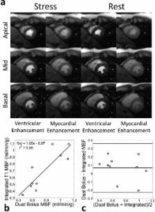
Figure 1
Comparison of proposed single-bolus non-ECG-triggered and conventional dual-bolus
ECG-triggered methods. (a) Comparison of example ECG-triggered Cartesian and the non-ECG-triggered
radial images in a rest perfusion study. Because ECG-triggered Cartesian images are
acquired sequentially, they fall during different cardiac phases (mid-systole in the
apical compared to early-diastole of the basal slice). (b) Correlation of mean MBF
found between the two techniques (r2 = 0.86). (b) A Bland-Altman plot comparing the
two methods.
Figure 2
Example non-ECG-triggered stress-rest perfusion images from a healthy volunteer study.
Each image was reconstructed from 26 projections. Stress images were acquired first.
All images were acquired during systole to maximize myocardial thickness. Images of
peak ventricular enhancement and peak myocardial enhancement are shown.