170
views
0
recommends
+1 Recommend
0 collections
    0
    shares
      • Record: found
      • Abstract: found
      • Article: found
      Is Open Access

      The efficacy of maxillary protraction protocols with the micro-implant-assisted rapid palatal expander (MARPE) and the novel N2 mini-implant—a finite element study

      research-article

      Read this article at

      Bookmark
          There is no author summary for this article yet. Authors can add summaries to their articles on ScienceOpen to make them more accessible to a non-specialist audience.

          Abstract

          Background

          Maxillary protraction with the novel N2 mini-implant- and micro-implant-assisted rapid palatal expander (MARPE) can potentially provide significant skeletal effects without surgery, even in older patients where conventional facemask therapy has limited skeletal effects. However, the skeletal effects of altering the location and direction of force from mini-implant-assisted maxillary protraction have not been extensively analyzed. In this study, the application of the novel N2 mini-implant as an orthopedic anchorage device is explored in its ability to treat patients with class III malocclusions.

          Methods

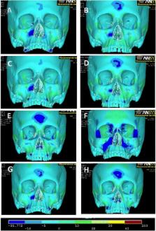
          A 3D cranial mesh model with associated sutures was developed from CT images and Mimics modeling software. Utilizing ANSYS simulation software, protraction forces were applied at different locations and directions to simulate conventional facemask therapy and seven maxillary protraction protocols utilizing the novel N2 mini-implant. Stress distribution and displacement were analyzed. Video animations and superimpositions were created.

          Results

          By changing the vector of force and location of N2 mini-implant, the maxilla was displaced differentially. Varying degrees of forward, downward, and rotational movements were observed in each case. For brachyfacial patients, anterior micro-implant-supported protraction at −45° or intermaxillary class III elastics at −45° are recommended. For dolicofacial patients, either anterior micro-implants at −15° or an intermaxillary spring at +30° is recommended. For mesofacial patients with favorable vertical maxillary position, palatal micro-implants at −30° are recommended; anterior micro-implants at −30° are preferred for shallow bites. For patients with a severe mid-facial deficiency, intermaxillary class III elastics at −30° are most effective in promoting anterior growth of the maxilla.

          Conclusions

          By varying the location of N2 mini-implants and vector of class III mechanics, clinicians can differentially alter the magnitude of forward, downward, and rotational movement of the maxilla. As a result, treatment protocol can be customized for each unique class III patient.

          Related collections

          Most cited references19

          • Record: found
          • Abstract: found
          • Article: not found

          Comparison of two protocols for maxillary protraction: bone anchors versus face mask with rapid maxillary expansion.

          To test the hypothesis that there is no difference in the active treatment effects for maxillary advancement induced by bone-anchored maxillary protraction (BAMP) and the active treatment effects for face mask in association with rapid maxillary expansion (RME/FM). This is a study on consecutively treated patients. The changes in dentoskeletal cephalometric variables from start of treatment (T1) to end of active treatment (T2) with an average T1-T2 interval of about 1 year were contrasted in a BAMP sample of 21 subjects with a RME/FM sample of 34 patients. All subjects were prepubertal at T1. Statistical comparison was performed with t-tests for independent samples. The BAMP protocol produced significantly larger maxillary advancement than the RME/FM therapy (with a difference of 2 mm to 3 mm). Mandibular sagittal changes were similar, while vertical changes were better controlled with BAMP. The sagittal intermaxillary relationships improved 2.5 mm more in the BAMP patients. Additional favorable outcomes of BAMP treatment were the lack of clockwise rotation of the mandible as well as a lack of retroclination of the lower incisors. The hypothesis is rejected. The BAMP protocol produced significantly larger maxillary advancement than the RME/FM therapy.
            Bookmark
            • Record: found
            • Abstract: found
            • Article: not found

            Application and effectiveness of a mini-implant- and tooth-borne rapid palatal expansion device: the hybrid hyrax.

            Rapid palatal expansion (RPE) is used for treatment of skeletal crossbites. It may be combined with a face mask if the maxilla is to be protracted. Conventional tooth-borne appliances rely on an almost complete dentition to transmit the relatively high forces to the bony structures of the maxilla and midface. In most situations, tooth-borne appliances produce adverse effects such as buccal tipping of the lateral teeth, imposing the risk of recessions and vestibular bone fenestrations. To overcome these drawbacks, an RPE appliance was developed that utilizes mini-implants anteriorly in the palate for skeletal anchorage. Because this device is also attached to the first molars, it can be denominated as a bone- and tooth-borne appliance (hybrid hyrax). The objective of this clinical pilot study was to investigate its dental and skeletal effects. RPE was performed in 13 patients (seven females, six males; mean age 11.2 years). In 10 patients with a skeletal Class III occlusion, a face mask was used simultaneously for maxillary protraction. Three-dimensional scans of the individual study models were digitally superimposed for the assessment of the dental effects. Skeletal effects were evaluated by lateral cephalograms taken before and after RPE and protraction. The time needed to achieve the intended expansion ranged from 4 to 14 days (mean 8.7 ± 3.6 days). The mean expansion in the first premolar/first primary molar region was 6.3 ± 2.9 mm and 5.0 ± 1.5 mm in the first molar region. The Wits appraisal changed from -5.2 ± 1.3 mm to -2.5 ± 1.5 mm (mean improvement 2.7 ± 1.3 mm). The right first molar migrated 0.4 ± 0.6 mm mesially and the left one 0.3 ± 0.2 mm. The hybrid hyrax is effective for RPE and can be employed especially in patients with reduced anterior dental anchorage. Since most teeth are not in the appliance, regular orthodontic treatment can start early. The combination of the hybrid hyrax with a face mask for maxillary protraction appears to be effective in minimizing mesial migration of the dentition. © 2010 BY QUINTESSENCE PUBLISHING CO, INC.
              Bookmark
              • Record: found
              • Abstract: found
              • Article: not found

              Treatment response and long-term dentofacial adaptations to maxillary expansion and protraction.

              The purpose of this article is to summarize the short-term and long-term results of the authors' clinical prospective study on the treatment of Class III malocclusion using the protraction facemask. An attempt is made to answer questions pertaining to this treatment modality. Twenty patients with skeletal Class III malocclusion were treated consecutively with maxillary expansion and a protraction facemask. A positive overjet was obtained in all cases after 6 to 9 months of treatment. These changes were contributed to by a forward movement of the maxilla, backward and downward rotation of the mandible, proclination of the maxillary incisors, and retroclination of the mandibular incisors. The molar relationship was overcorrected to Class I or Class II dental arch relationship. The overbite was reduced with a significant increase in lower facial height. The treatment was found to be stable 2 years after removal of the appliances. At the end of the 4-year observation period, 15 of the 20 patients maintained a positive overjet or an end-to-end incisal relationship. Patients who reverted back to a negative overjet were found to have excess horizontal mandibular growth that was not compensated by proclination of the maxillary incisors. A review of the literature showed that maxillary expansion in conjunction with protraction produced greater forward movement of the maxilla. Maxillary protraction with a 30 degrees forward and downward force applied at the canine region produced an acceptable clinical response. The reciprocal force from maxillary protraction transmitted to the temporomandibular joint did not increase masticatory muscle pain or activity. Significant soft tissue profile change can be expected with maxillary protraction including straightening of the facial profile and better lip competence and posture. However, one should anticipate individual variations in treatment response and subsequent growth changes. Treatment with the protraction facemask is most effective in Class III patients with a retrusive maxilla and a hypodivergent growth pattern. Treatment initiated at the time of initial eruption of the upper central incisors helps to maintain the anterior occlusion after treatment.
                Bookmark

                Author and article information

                Contributors
                wmoon@dentistry.ucla.edu
                kimberley.wu@gmail.com
                macginnis@gmail.com
                high5jay@gmail.com
                howard.dmd@gmail.com
                gyoussef@csun.edu
                awmachado@bol.com.br
                Journal
                Prog Orthod
                Prog Orthod
                Progress in Orthodontics
                Springer Berlin Heidelberg (Berlin/Heidelberg )
                1723-7785
                2196-1042
                4 June 2015
                4 June 2015
                2015
                : 16
                : 16
                Affiliations
                [ ]UCLA Section of Orthodontics, UCLA School of Dentistry, 10833 Le Conte Avenue, CHS – Box 951668, Los Angeles, CA 90095-1668 USA
                [ ]Mechanical Engineering Department, California State University, Northridge, 18111 Nordhoff Street, Northridge, CA 91330 USA
                [ ]Section of Orthodontics, Federal University of Bahia, Salvador, Bahia Brazil
                Article
                83
                10.1186/s40510-015-0083-z
                4456601
                26061987
                cac16375-dd2c-40f9-baf5-3fe2a17d369b
                © Moon et al.; licensee Springer. 2015

                This is an open access article distributed under the terms of the Creative Commons Attribution License ( http://creativecommons.org/licenses/by/4.0), which permits unrestricted use, distribution, and reproduction in any medium, provided the original work is properly credited.

                History
                : 4 March 2015
                : 27 April 2015
                Categories
                Research
                Custom metadata
                © The Author(s) 2015

                Comments

                Comment on this article