16
views
0
recommends
+1 Recommend
0 collections
    0
    shares
      • Record: found
      • Abstract: found
      • Article: found
      Is Open Access

      Electrocardiographic methods for diagnosis and risk stratification in the Brugada syndrome

      review-article
      a , b , b , b , *
      Journal of the Saudi Heart Association
      Elsevier
      AP, action potential, ARI, activation-recovery intervals, BrS, Brugada syndrome, ECG, electrocardiogram, EPS, electrophysiology study, ICD, implantable cardioverter-defibrillator, IHD, ischaemic heart disease, LBBB, left bundle branch block, MAP, monophasic action potential, MI, myocardial infarction, PCA, principal component analysis, RVOT, right ventricular outflow tract, SAECG, signal-averaged electrocardiogram, SCD, sudden cardiac death, SNP, single-nucleotide polymorphism, VF, ventricular fibrillation, VT, ventricular tachycardia, WT, wavelet transform, Brugada syndrome, Electrocardiogram, Sudden cardiac death, Risk stratification, Genetic arrhythmic syndromes

      Read this article at

      Bookmark
          There is no author summary for this article yet. Authors can add summaries to their articles on ScienceOpen to make them more accessible to a non-specialist audience.

          Abstract

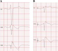
          The Brugada syndrome (BrS) is a malignant, genetically-determined, arrhythmic syndrome manifesting as syncope or sudden cardiac death (SCD) in individuals with structurally normal hearts. The diagnosis of the BrS is mainly based on the presence of a spontaneous or Na + channel blocker induced characteristic, electrocardiographic (ECG) pattern (type 1 or coved Brugada ECG pattern) typically seen in leads V1 and V2 recorded from the 4th to 2nd intercostal (i.c.) spaces. This pattern needs to be distinguished from similar ECG changes due to other causes (Brugada ECG phenocopies). This review focuses mainly on the ECG-based methods for diagnosis and arrhythmia risk assessment in the BrS. Presently, the main unresolved clinical problem is the identification of those patients at high risk of SCD who need implantable cardioverter-defibrillator (ICD), which is the only therapy with proven efficacy. Current guidelines recommend ICD implantation only in patients with spontaneous type 1 ECG pattern, and either history of aborted cardiac arrest or documented sustained VT (class I), or syncope of arrhythmic origin (class IIa) because they are at high risk of recurrent arrhythmic events (up to 10% or more annually for those with aborted cardiac arrest). The majority of BrS patients are asymptomatic when diagnosed and considered to have low risk (around 0.5% annually) and therefore not indicated for ICD. The majority of SCD victims in the BrS, however, had no symptoms prior to the fatal event and therefore were not protected with an ICD. While some ECG markers such as QRS fragmentation, infero-lateral early repolarisation, and abnormal late potentials on signal-averaged ECG are known to be linked to increased arrhythmic risk, they are not sufficiently sensitive or specific. Potential novel ECG-based strategies for risk stratification are discussed based on computerised methods for depolarisation and repolarisation analysis, a composite approach targeting several major components of ventricular arrhythmogenesis, and the collection of large digital ECG databases in genotyped BrS patients and their relatives.

          Related collections

          Most cited references89

          • Record: found
          • Abstract: found
          • Article: not found

          Right bundle branch block, persistent ST segment elevation and sudden cardiac death: a distinct clinical and electrocardiographic syndrome. A multicenter report.

          The objectives of this study were to present data on eight patients with recurrent episodes of aborted sudden death unexplainable by currently known diseases whose common clinical and electrocardiographic (ECG) features define them as having a distinct syndrome different from idiopathic ventricular fibrillation. Among patients with ventricular arrhythmias who have no structural heart disease, several subgroups have been defined. The present patients constitute an additional subgroup with these findings. The study group consisted of eight patients, six male and two female, with recurrent episodes of aborted sudden death. Clinical and laboratory data and results of electrocardiography, electrophysiology, echocardiography, angiography, histologic study and exercise testing were available in most cases. The ECG during sinus rhythm showed right bundle branch block, normal QT interval and persistent ST segment elevation in precordial leads V1 to V2-V3 not explainable by electrolyte disturbances, ischemia or structural heart disease. No histologic abnormalities were found in the four patients in whom ventricular biopsies were performed. The arrhythmia leading to (aborted) sudden death was a rapid polymorphic ventricular tachycardia initiating after a short coupled ventricular extrasystole. A similar arrhythmia was initiated by two to three ventricular extrastimuli in four of the seven patients studied by programmed electrical stimulation. Four patients had a prolonged HV interval during sinus rhythm. One patient receiving amiodarone died suddenly during implantation of a demand ventricular pacemaker. The arrhythmia of two patients was controlled with a beta-adrenergic blocking agent. Four patients received an implantable defibrillator that was subsequently used by one of them, and all four are alive. The remaining patient received a demand ventricular pacemaker and his arrhythmia is controlled with amiodarone and diphenylhydantoin. Common clinical and ECG features define a distinct syndrome in this group of patients. Its causes remain unknown.
            Bookmark
            • Record: found
            • Abstract: found
            • Article: not found

            Long-term prognosis of patients diagnosed with Brugada syndrome: Results from the FINGER Brugada Syndrome Registry.

            Brugada syndrome is characterized by ST-segment elevation in the right precordial leads and an increased risk of sudden cardiac death (SCD). Fundamental questions remain on the best strategy for assessing the real disease-associated arrhythmic risk, especially in asymptomatic patients. The aim of the present study was to evaluate the prognosis and risk factors of SCD in Brugada syndrome patients in the FINGER (France, Italy, Netherlands, Germany) Brugada syndrome registry. Patients were recruited in 11 tertiary centers in 4 European countries. Inclusion criteria consisted of a type 1 ECG present either at baseline or after drug challenge, after exclusion of diseases that mimic Brugada syndrome. The registry included 1029 consecutive individuals (745 men; 72%) with a median age of 45 (35 to 55) years. Diagnosis was based on (1) aborted SCD (6%); (2) syncope, otherwise unexplained (30%); and (3) asymptomatic patients (64%). During a median follow-up of 31.9 (14 to 54.4) months, 51 cardiac events (5%) occurred (44 patients experienced appropriate implantable cardioverter-defibrillator shocks, and 7 died suddenly). The cardiac event rate per year was 7.7% in patients with aborted SCD, 1.9% in patients with syncope, and 0.5% in asymptomatic patients. Symptoms and spontaneous type 1 ECG were predictors of arrhythmic events, whereas gender, familial history of SCD, inducibility of ventricular tachyarrhythmias during electrophysiological study, and the presence of an SCN5A mutation were not predictive of arrhythmic events. In the largest series of Brugada syndrome patients thus far, event rates in asymptomatic patients were low. Inducibility of ventricular tachyarrhythmia and family history of SCD were not predictors of cardiac events.
              Bookmark
              • Record: found
              • Abstract: found
              • Article: not found

              Cellular basis for the Brugada syndrome and other mechanisms of arrhythmogenesis associated with ST-segment elevation.

              The Brugada syndrome is characterized by marked ST-segment elevation in the right precordial ECG leads and is associated with a high incidence of sudden and unexpected arrhythmic death. Our study examines the cellular basis for this syndrome. Using arterially perfused wedges of canine right ventricle (RV), we simultaneously recorded transmembrane action potentials from 2 epicardial and 1 endocardial sites, together with unipolar electrograms and a transmural ECG. Loss of the action potential dome in epicardium but not endocardium after exposure to pinacidil (2 to 5 micromol/L), a K(+) channel opener, or the combination of a Na(+) channel blocker (flecainide, 7 micromol/L) and acetylcholine (ACh, 2 to 3 micromol/L) resulted in an abbreviation of epicardial response and a transmural dispersion of repolarization, which caused an ST-segment elevation in the ECG. ACh facilitated loss of the action potential dome, whereas isoproterenol (0.1 to 1 micromol/L) restored the epicardial dome, thus reducing or eliminating the ST-segment elevation. Heterogeneous loss of the dome caused a marked dispersion of repolarization within the epicardium and transmurally, thus giving rise to phase 2 reentrant extrasystole, which precipitated ventricular tachycardia (VT) and ventricular fibrillation (VF). Transient outward current (I(to)) block with 4-aminopyridine (1 to 2 mmol/L) or quinidine (5 micromol/L) restored the dome, normalized the ST segment, and prevented VT/VF. Conclusions-Depression or loss of the action potential dome in RV epicardium creates a transmural voltage gradient that may be responsible for the ST-segment elevation observed in the Brugada syndrome and other syndromes exhibiting similar ECG manifestations. Our results also demonstrate that extrasystolic activity due to phase 2 reentry can arise in the intact wall of the canine RV and serve as the trigger for VT/VF. Our data point to I(to) block (4-aminopyridine, quinidine) as an effective pharmacological treatment.
                Bookmark

                Author and article information

                Contributors
                Journal
                J Saudi Heart Assoc
                J Saudi Heart Assoc
                Journal of the Saudi Heart Association
                Elsevier
                1016-7315
                2212-5043
                03 July 2014
                April 2015
                03 July 2014
                : 27
                : 2
                : 96-108
                Affiliations
                [a ]Center for Health Studies, Prince Sultan Military Medical City, Riyadh, Saudi Arabia
                [b ]Cardiac and Vascular Sciences Research Centre, St. George’s University of London, London, United Kingdom
                Author notes
                [* ]Corresponding author. Address: Cardiac and Vascular Sciences Research Centre, St. George’s University of London Cranmer Terrace, London SW17 0RE, United Kingdom. Tel.: +44 (0)208 725 3708. vbatchva@ 123456sgul.ac.uk
                Article
                S1016-7315(14)00069-4
                10.1016/j.jsha.2014.06.004
                4392351
                25870503
                cb2d8d2d-9d32-47c8-b9b2-15817f06d34d
                © 2014 Production and hosting by Elsevier B.V. on behalf of King Saud University.

                This is an open access article under the CC BY-NC-ND license (http://creativecommons.org/licenses/by-nc-nd/3.0/).

                History
                : 1 May 2014
                : 2 June 2014
                : 26 June 2014
                Categories
                Review Article

                ap, action potential,ari, activation-recovery intervals,brs, brugada syndrome,ecg, electrocardiogram,eps, electrophysiology study,icd, implantable cardioverter-defibrillator,ihd, ischaemic heart disease,lbbb, left bundle branch block,map, monophasic action potential,mi, myocardial infarction,pca, principal component analysis,rvot, right ventricular outflow tract,saecg, signal-averaged electrocardiogram,scd, sudden cardiac death,snp, single-nucleotide polymorphism,vf, ventricular fibrillation,vt, ventricular tachycardia,wt, wavelet transform,brugada syndrome,electrocardiogram,sudden cardiac death,risk stratification,genetic arrhythmic syndromes

                Comments

                Comment on this article