39
views
0
recommends
+1 Recommend
0 collections
    0
    shares
      • Record: found
      • Abstract: found
      • Article: found
      Is Open Access

      Textural features in pre-treatment [F18]-FDG-PET/CT are correlated with risk of local recurrence and disease-specific survival in early stage NSCLC patients receiving primary stereotactic radiation therapy

      research-article

      Read this article at

      Bookmark
          There is no author summary for this article yet. Authors can add summaries to their articles on ScienceOpen to make them more accessible to a non-specialist audience.

          Abstract

          Background

          Textural features in FDG-PET have been shown to provide prognostic information in a variety of tumor entities. Here we evaluate their predictive value for recurrence and prognosis in NSCLC patients receiving primary stereotactic radiation therapy (SBRT).

          Methods

          45 patients with early stage NSCLC (T1 or T2 tumor, no lymph node or distant metastases) were included in this retrospective study and followed over a median of 21.4 months (range 3.1–71.1). All patients were considered non-operable due to concomitant disease and referred to SBRT as the primary treatment modality. Pre-treatment FDG-PET/CT scans were obtained from all patients. SUV and volume-based analysis as well as extraction of textural features based on neighborhood gray-tone difference matrices (NGTDM) and gray-level co-occurence matrices (GLCM) were performed using InterView Fusion™ (Mediso Inc., Budapest). The ability to predict local recurrence (LR), lymph node (LN) and distant metastases (DM) was measured using the receiver operating characteristic (ROC). Univariate and multivariate analysis of overall and disease-specific survival were executed.

          Results

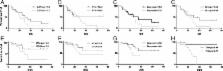
          7 out of 45 patients (16%) experienced LR, 11 (24%) LN and 11 (24%) DM. ROC revealed a significant correlation of several textural parameters with LR with an AUC value for entropy of 0.872. While there was also a significant correlation of LR with tumor size in the overall cohort, only texture was predictive when examining T1 (tumor diameter < = 3 cm) and T2 (>3 cm) subgroups. No correlation of the examined PET parameters with LN or DM was shown.

          In univariate survival analysis, both heterogeneity and tumor size were predictive for disease-specific survival, but only texture determined by entropy was determined as an independent factor in multivariate analysis (hazard ratio 7.48, p = .016). Overall survival was not significantly correlated to any examined parameter, most likely due to the high comorbidity in our cohort.

          Conclusions

          Our study adds to the growing evidence that tumor heterogeneity as described by FDG-PET texture is associated with response to radiation therapy in NSCLC. The results may be helpful into identifying patients who might profit from an intensified treatment regime, but need to be verified in a prospective patient cohort before being incorporated into routine clinical practice.

          Related collections

          Most cited references27

          • Record: found
          • Abstract: found
          • Article: not found

          Intratumor heterogeneity characterized by textural features on baseline 18F-FDG PET images predicts response to concomitant radiochemotherapy in esophageal cancer.

          (18)F-FDG PET is often used in clinical routine for diagnosis, staging, and response to therapy assessment or prediction. The standardized uptake value (SUV) in the primary or regional area is the most common quantitative measurement derived from PET images used for those purposes. The aim of this study was to propose and evaluate new parameters obtained by textural analysis of baseline PET scans for the prediction of therapy response in esophageal cancer. Forty-one patients with newly diagnosed esophageal cancer treated with combined radiochemotherapy were included in this study. All patients underwent pretreatment whole-body (18)F-FDG PET. Patients were treated with radiotherapy and alkylatinlike agents (5-fluorouracil-cisplatin or 5-fluorouracil-carboplatin). Patients were classified as nonresponders (progressive or stable disease), partial responders, or complete responders according to the Response Evaluation Criteria in Solid Tumors. Different image-derived indices obtained from the pretreatment PET tumor images were considered. These included usual indices such as maximum SUV, peak SUV, and mean SUV and a total of 38 features (such as entropy, size, and magnitude of local and global heterogeneous and homogeneous tumor regions) extracted from the 5 different textures considered. The capacity of each parameter to classify patients with respect to response to therapy was assessed using the Kruskal-Wallis test (P < 0.05). Specificity and sensitivity (including 95% confidence intervals) for each of the studied parameters were derived using receiver-operating-characteristic curves. Relationships between pairs of voxels, characterizing local tumor metabolic nonuniformities, were able to significantly differentiate all 3 patient groups (P < 0.0006). Regional measures of tumor characteristics, such as size of nonuniform metabolic regions and corresponding intensity nonuniformities within these regions, were also significant factors for prediction of response to therapy (P = 0.0002). Receiver-operating-characteristic curve analysis showed that tumor textural analysis can provide nonresponder, partial-responder, and complete-responder patient identification with higher sensitivity (76%-92%) than any SUV measurement. Textural features of tumor metabolic distribution extracted from baseline (18)F-FDG PET images allow for the best stratification of esophageal carcinoma patients in the context of therapy-response prediction.
            Bookmark
            • Record: found
            • Abstract: found
            • Article: not found

            Tumor texture analysis in 18F-FDG PET: relationships between texture parameters, histogram indices, standardized uptake values, metabolic volumes, and total lesion glycolysis.

            Texture indices are of growing interest for tumor characterization in (18)F-FDG PET. Yet, on the basis of results published in the literature so far, it is unclear which indices should be used, what they represent, and how they relate to conventional indices such as standardized uptake values (SUVs), metabolic volume (MV), and total lesion glycolysis (TLG). We investigated in detail 31 texture indices, 5 first-order statistics (histogram indices) derived from the gray-level histogram of the tumor region, and their relationship with SUV, MV, and TLG in 3 different tumor types. Three patient groups corresponding to 3 cancer types at baseline were studied independently: patients with metastatic colorectal cancer (72 lesions), non-small cell lung cancer (24 lesions), and breast cancer (54 lesions). Thirty-one texture indices were studied in addition to SUVs, MV, and TLG, and 5 indices extracted from histogram analysis were also investigated. The relationships between indices were studied as well as the robustness of the various texture indices with respect to the parameters involved in the calculation method (sampling schemes and tumor volume of interest). Regardless of the patient group, many indices were highly correlated (Pearson correlation coefficient |r| ≥ 0.80), making it desirable to focus on only a few uncorrelated indices. Three histogram indices were highly correlated with SUVs (|r| ≥ 0.84). Four texture indices were highly correlated with MV, and none was highly correlated with SUVs (|r| ≤ 0.55). The resampling formula used to calculate texture indices had a substantial impact, and resampling using at least 32 discrete values should be used for texture indices calculation. The texture indices change as a function of the segmentation method was higher than that of peak and maximum SUVs but less than mean SUV for 5 texture indices and was larger than that of MV for 14 texture indices and for the 5 histogram indices. All these results were extremely consistent across the 3 tumor types and explained many of the observations reported in the literature so far. None of the histogram indices and only 17 of 31 texture indices were robust with respect to the tumor-segmentation method. An appropriate resampling formula with at least 32 gray levels should be used to avoid introducing a misleading relationship between texture indices and SUV. Some texture indices are highly correlated or strongly correlate with MV whatever the tumor type. Such correlation should be accounted for when interpreting the usefulness of texture indices for tumor characterization, which might call for systematic multivariate analyses.
              Bookmark
              • Record: found
              • Abstract: found
              • Article: not found

              Reproducibility of tumor uptake heterogeneity characterization through textural feature analysis in 18F-FDG PET.

              (18)F-FDG PET measurement of standardized uptake value (SUV) is increasingly used for monitoring therapy response and predicting outcome. Alternative parameters computed through textural analysis were recently proposed to quantify the heterogeneity of tracer uptake by tumors as a significant predictor of response. The primary objective of this study was to evaluate the reproducibility of these heterogeneity measurements. Double baseline (18)F-FDG PET scans were acquired within 4 d of each other for 16 patients before any treatment was considered. A Bland-Altman analysis was performed on 8 parameters based on histogram measurements and 17 parameters based on textural heterogeneity features after discretization with values between 8 and 128. The reproducibility of maximum and mean SUV was similar to that in previously reported studies, with a mean percentage difference of 4.7% ± 19.5% and 5.5% ± 21.2%, respectively. By comparison, better reproducibility was measured for some textural features describing local heterogeneity of tracer uptake, such as entropy and homogeneity, with a mean percentage difference of -2% ± 5.4% and 1.8% ± 11.5%, respectively. Several regional heterogeneity parameters such as variability in the intensity and size of regions of homogeneous activity distribution had reproducibility similar to that of SUV measurements, with 95% confidence intervals of -22.5% to 3.1% and -1.1% to 23.5%, respectively. These parameters were largely insensitive to the discretization range. Several parameters derived from textural analysis describing heterogeneity of tracer uptake by tumors on local and regional scales had reproducibility similar to or better than that of simple SUV measurements. These reproducibility results suggest that these (18)F-FDG PET-derived parameters, which have already been shown to have predictive and prognostic value in certain cancer models, may be used to monitor therapy response and predict patient outcome.
                Bookmark

                Author and article information

                Contributors
                thomas.pyka@tum.de
                ralph.bundschuh@ukb.uni-bonn.de
                Nicolaus.Andratschke@usz.ch
                wellenspiel@gmx.net
                Hanno.Specht@mri.tum.de
                laszlo.papp@mediso.com
                norbert.zsoter@mediso.hu
                markus.essler@ukb.uni-bonn.de
                Journal
                Radiat Oncol
                Radiat Oncol
                Radiation Oncology (London, England)
                BioMed Central (London )
                1748-717X
                22 April 2015
                22 April 2015
                2015
                : 10
                : 100
                Affiliations
                [ ]Nuklearmedizinische Klinik und Poliklinik, Klinikum rechts der Isar der TU München, Ismaninger Str, Munich, Germany
                [ ]Klinik und Poliklinik für Nuklearmedizin, Rheinische Friedrich-Wilhelms-Universität Bonn, Sigmund-Freud-Straße, Bonn, Germany
                [ ]Klinik für Strahlentherapie und Radiologische Onkologie, Klinikum rechts der Isar der TU München, Ismaninger Str, Munich, Germany
                [ ]Klinik für Radio-Onkologie, UniversitätsSpital Zürich, Rämistrasse, Zurich, Switzerland
                [ ]Mediso Medical Imaging Systems, Alsotorokvesz, Budapest, Hungary
                Article
                407
                10.1186/s13014-015-0407-7
                4465163
                25900186
                cc7fac78-d39b-4867-a13a-fd35307f8bd6
                © Pyka et al.; licensee BioMed Central. 2015

                This is an Open Access article distributed under the terms of the Creative Commons Attribution License ( http://creativecommons.org/licenses/by/4.0), which permits unrestricted use, distribution, and reproduction in any medium, provided the original work is properly credited. The Creative Commons Public Domain Dedication waiver ( http://creativecommons.org/publicdomain/zero/1.0/) applies to the data made available in this article, unless otherwise stated.

                History
                : 25 November 2014
                : 13 April 2015
                Categories
                Research
                Custom metadata
                © The Author(s) 2015

                Oncology & Radiotherapy
                stereotactic body radiation therapy,nsclc,fdg-pet,textural analysis
                Oncology & Radiotherapy
                stereotactic body radiation therapy, nsclc, fdg-pet, textural analysis

                Comments

                Comment on this article