9
views
0
recommends
+1 Recommend
0 collections
    0
    shares
      • Record: found
      • Abstract: found
      • Article: found
      Is Open Access

      Performance of HADDOCK and a simple contact-based protein–ligand binding affinity predictor in the D3R Grand Challenge 2

      research-article

      Read this article at

      Bookmark
          There is no author summary for this article yet. Authors can add summaries to their articles on ScienceOpen to make them more accessible to a non-specialist audience.

          Abstract

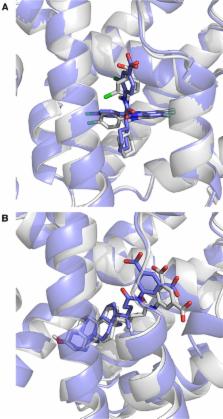
          We present the performance of HADDOCK, our information-driven docking software, in the second edition of the D3R Grand Challenge. In this blind experiment, participants were requested to predict the structures and binding affinities of complexes between the Farnesoid X nuclear receptor and 102 different ligands. The models obtained in Stage1 with HADDOCK and ligand-specific protocol show an average ligand RMSD of 5.1 Å from the crystal structure. Only 6/35 targets were within 2.5 Å RMSD from the reference, which prompted us to investigate the limiting factors and revise our protocol for Stage2. The choice of the receptor conformation appeared to have the strongest influence on the results. Our Stage2 models were of higher quality (13 out of 35 were within 2.5 Å), with an average RMSD of 4.1 Å. The docking protocol was applied to all 102 ligands to generate poses for binding affinity prediction. We developed a modified version of our contact-based binding affinity predictor PRODIGY, using the number of interatomic contacts classified by their type and the intermolecular electrostatic energy. This simple structure-based binding affinity predictor shows a Kendall’s Tau correlation of 0.37 in ranking the ligands (7th best out of 77 methods, 5th/25 groups). Those results were obtained from the average prediction over the top10 poses, irrespective of their similarity/correctness, underscoring the robustness of our simple predictor. This results in an enrichment factor of 2.5 compared to a random predictor for ranking ligands within the top 25%, making it a promising approach to identify lead compounds in virtual screening.

          Electronic supplementary material

          The online version of this article (doi:10.1007/s10822-017-0049-y) contains supplementary material, which is available to authorized users.

          Related collections

          Most cited references30

          • Record: found
          • Abstract: found
          • Article: found
          Is Open Access

          BindingDB in 2015: A public database for medicinal chemistry, computational chemistry and systems pharmacology

          BindingDB, www.bindingdb.org, is a publicly accessible database of experimental protein-small molecule interaction data. Its collection of over a million data entries derives primarily from scientific articles and, increasingly, US patents. BindingDB provides many ways to browse and search for data of interest, including an advanced search tool, which can cross searches of multiple query types, including text, chemical structure, protein sequence and numerical affinities. The PDB and PubMed provide links to data in BindingDB, and vice versa; and BindingDB provides links to pathway information, the ZINC catalog of available compounds, and other resources. The BindingDB website offers specialized tools that take advantage of its large data collection, including ones to generate hypotheses for the protein targets bound by a bioactive compound, and for the compounds bound by a new protein of known sequence; and virtual compound screening by maximal chemical similarity, binary kernel discrimination, and support vector machine methods. Specialized data sets are also available, such as binding data for hundreds of congeneric series of ligands, drawn from BindingDB and organized for use in validating drug design methods. BindingDB offers several forms of programmatic access, and comes with extensive background material and documentation. Here, we provide the first update of BindingDB since 2007, focusing on new and unique features and highlighting directions of importance to the field as a whole.
            Bookmark
            • Record: found
            • Abstract: found
            • Article: not found

            MMTSB Tool Set: enhanced sampling and multiscale modeling methods for applications in structural biology.

            We describe the Multiscale Modeling Tools for Structural Biology (MMTSB) Tool Set (https://mmtsb.scripps.edu/software/mmtsbToolSet.html), which is a novel set of utilities and programming libraries that provide new enhanced sampling and multiscale modeling techniques for the simulation of proteins and nucleic acids. The tool set interfaces with the existing molecular modeling packages CHARMM and Amber for classical all-atom simulations, and with MONSSTER for lattice-based low-resolution conformational sampling. In addition, it adds new functionality for the integration and translation between both levels of detail. The replica exchange method is implemented to allow enhanced sampling of both the all-atom and low-resolution models. The tool set aims at applications in structural biology that involve protein or nucleic acid structure prediction, refinement, and/or extended conformational sampling. With structure prediction applications in mind, the tool set also implements a facility that allows the control and application of modeling tasks on a large set of conformations in what we have termed ensemble computing. Ensemble computing encompasses loosely coupled, parallel computation on high-end parallel computers, clustered computational grids and desktop grid environments. This paper describes the design and implementation of the MMTSB Tool Set and illustrates its utility with three typical examples--scoring of a set of predicted protein conformations in order to identify the most native-like structures, ab initio folding of peptides in implicit solvent with the replica exchange method, and the prediction of a missing fragment in a larger protein structure.
              Bookmark
              • Record: found
              • Abstract: found
              • Article: not found

              Plectasin, a fungal defensin, targets the bacterial cell wall precursor Lipid II.

              Host defense peptides such as defensins are components of innate immunity and have retained antibiotic activity throughout evolution. Their activity is thought to be due to amphipathic structures, which enable binding and disruption of microbial cytoplasmic membranes. Contrary to this, we show that plectasin, a fungal defensin, acts by directly binding the bacterial cell-wall precursor Lipid II. A wide range of genetic and biochemical approaches identify cell-wall biosynthesis as the pathway targeted by plectasin. In vitro assays for cell-wall synthesis identified Lipid II as the specific cellular target. Consistently, binding studies confirmed the formation of an equimolar stoichiometric complex between Lipid II and plectasin. Furthermore, key residues in plectasin involved in complex formation were identified using nuclear magnetic resonance spectroscopy and computational modeling.
                Bookmark

                Author and article information

                Contributors
                +31 30 2533859 , a.m.j.j.bonvin@uu.nl
                Journal
                J Comput Aided Mol Des
                J. Comput. Aided Mol. Des
                Journal of Computer-Aided Molecular Design
                Springer International Publishing (Cham )
                0920-654X
                1573-4951
                22 August 2017
                22 August 2017
                2018
                : 32
                : 1
                : 175-185
                Affiliations
                [1 ]ISNI 0000000120346234, GRID grid.5477.1, Bijvoet Center for Biomolecular Research, Faculty of Science - Chemistry, , Utrecht University, ; Padualaan 8, 3584CH Utrecht, The Netherlands
                [2 ]ISNI 0000 0000 9511 4342, GRID grid.8051.c, CNC - Center for Neuroscience and Cell Biology, FMUC, , Universidade de Coimbra, ; Rua Larga, Polo I, 1ºandar, 3004-517 Coimbra, Portugal
                [3 ]ISNI 0000000419368956, GRID grid.168010.e, James H. Clark Center, , Stanford University, ; 318 Campus Drive, S210, Stanford, CA 94305 USA
                Author information
                http://orcid.org/0000-0001-7369-1322
                Article
                49
                10.1007/s10822-017-0049-y
                5767195
                28831657
                cd7c3909-86a3-4e20-ad68-f6c627e40862
                © The Author(s) 2017

                Open AccessThis article is distributed under the terms of the Creative Commons Attribution 4.0 International License ( http://creativecommons.org/licenses/by/4.0/), which permits unrestricted use, distribution, and reproduction in any medium, provided you give appropriate credit to the original author(s) and the source, provide a link to the Creative Commons license, and indicate if changes were made.

                History
                : 2 June 2017
                : 18 August 2017
                Funding
                Funded by: FundRef http://dx.doi.org/10.13039/501100003246, Nederlandse Organisatie voor Wetenschappelijk Onderzoek;
                Award ID: TOP-PUNT 718.015.001
                Award ID: VENI 722.014.005
                Award Recipient :
                Funded by: FundRef http://dx.doi.org/10.13039/100010661, Horizon 2020 Framework Programme;
                Award ID: West-Life grant no. 675858
                Award ID: BioExcel grant no. 675728
                Award ID: INDIGO-Datacloud grant no 653549
                Award Recipient :
                Funded by: FundRef http://dx.doi.org/10.13039/100010665, H2020 Marie Skłodowska-Curie Actions;
                Award ID: BAP-659025
                Award ID: MEMBRANEPROT-659826
                Award Recipient :
                Categories
                Article
                Custom metadata
                © Springer International Publishing AG, part of Springer Nature 2018

                Biomedical engineering
                d3r,drug design data resource,docking,binding affinity,ranking,intermolecular contacts

                Comments

                Comment on this article