13
views
0
recommends
+1 Recommend
0 collections
    0
    shares
      • Record: found
      • Abstract: found
      • Article: found
      Is Open Access

      Epidemiology and Pathophysiology of Glomerular C4d Staining in Native Kidney Biopsies

      research-article

      Read this article at

      Bookmark
          There is no author summary for this article yet. Authors can add summaries to their articles on ScienceOpen to make them more accessible to a non-specialist audience.

          Abstract

          Introduction

          Routine C4d staining in renal transplantation has stimulated its use in kidney biopsies with glomerulonephritis (GN). Methodical description on staining patterns in the native kidney is not available.

          Methods

          We retrospectively evaluated C4d staining in formalin-fixed paraffin-embedded sections from 519 native kidney biopsies (bx) with and without glomerular disease.

          Results

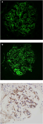
          Strong C4d staining was consistently present in immune-complex GN, including lupus nephritis (LN) ( n = 68), membranous GN ( n = 24), membranoproliferative glomerulonephritis (MPGN) pattern ( n = 22), fibrillary GN ( n = 3), and proliferative GN with monoclonal IgG ( n = 3). C4d stained all cases of postinfectious GN ( n = 7) amyloidosis ( n = 20) and C1q GN ( n = 3). In contrast, IgA nephropathy (IgAN) ( n = 34), was negative in 62% of bx, with the rest staining variably. The E1 Oxford classification score correlated with capillary wall C4d staining ( P = 0.05). C4d marked the glomerular and arteriolar lesions in thrombotic microangiopathy (TMA; n = 16), the glomerular sclerotic segments in focal segmental glomerulosclerosis (FSGS; n = 77), and marked areas of necrosis in crescentic GN ( n = 21). In diabetic glomerulopathy ( n = 70), C4d marked advanced insudative lesions but was negative otherwise. C4d weakly stained the mesangium, or was negative in normal biopsies ( n = 13), minimal change disease (MCD; n = 21), thin basement membrane disease ( n = 20), Alport ( n = 3), IgM nephropathy ( n = 2), C3 glomerulopathy ( n = 5), acute interstitial nephritis ( n = 12), acute tubular necrosis ( n = 22), ischemic glomerulopathy/nephrosclerosis ( n = 23), and other miscellaneous processes ( n = 14). Staining in tubular basement membranes and peritubular capillaries was most common in lupus.

          Conclusion

          Based on reliable staining in lupus and membranous GN, C4d staining is potentially useful as a screening and diagnostic tool, if only paraffin-embedded tissue is available. Knowledge of C4d staining patterns in normal and pathological tissues enhances its diagnostic value.

          Graphical abstract

          Related collections

          Most cited references69

          • Record: found
          • Abstract: found
          • Article: not found

          Pathologic classification of diabetic nephropathy.

          Although pathologic classifications exist for several renal diseases, including IgA nephropathy, focal segmental glomerulosclerosis, and lupus nephritis, a uniform classification for diabetic nephropathy is lacking. Our aim, commissioned by the Research Committee of the Renal Pathology Society, was to develop a consensus classification combining type1 and type 2 diabetic nephropathies. Such a classification should discriminate lesions by various degrees of severity that would be easy to use internationally in clinical practice. We divide diabetic nephropathy into four hierarchical glomerular lesions with a separate evaluation for degrees of interstitial and vascular involvement. Biopsies diagnosed as diabetic nephropathy are classified as follows: Class I, glomerular basement membrane thickening: isolated glomerular basement membrane thickening and only mild, nonspecific changes by light microscopy that do not meet the criteria of classes II through IV. Class II, mesangial expansion, mild (IIa) or severe (IIb): glomeruli classified as mild or severe mesangial expansion but without nodular sclerosis (Kimmelstiel-Wilson lesions) or global glomerulosclerosis in more than 50% of glomeruli. Class III, nodular sclerosis (Kimmelstiel-Wilson lesions): at least one glomerulus with nodular increase in mesangial matrix (Kimmelstiel-Wilson) without changes described in class IV. Class IV, advanced diabetic glomerulosclerosis: more than 50% global glomerulosclerosis with other clinical or pathologic evidence that sclerosis is attributable to diabetic nephropathy. A good interobserver reproducibility for the four classes of DN was shown (intraclass correlation coefficient = 0.84) in a test of this classification.
            Bookmark
            • Record: found
            • Abstract: found
            • Article: not found

            The role of complement in inflammatory diseases from behind the scenes into the spotlight.

            Our understanding of the biology of the complement system has undergone a drastic metamorphosis since its original discovery. This system, which was traditionally primarily described as a "complement" to humoral immunity, is now perceived as a central constituent of innate immunity, defending the host against pathogens, coordinating various events during inflammation, and bridging innate and adaptive immune responses. Complement is an assembly of proteins found in the blood and body fluids and on cell surfaces. Soluble complement components form the proteolytic cascade, whose activation leads to the generation of complement effectors that target various cells involved in the immune response. Membrane-bound receptors and regulators transmit signals from complement effectors to target cells and limit complement activation to the surfaces of pathogens and damaged or activated host cells. The multiple interconnections among complement proteins, immune cells, and mediators provide an excellent mechanism to protect the organism against infections and support the repair of damaged tissues. However, disturbances in this "defense machinery" contribute to the pathogenesis of various diseases. The role of complement in various inflammatory disorders is multifaceted; for example, the activation of complement can significantly contribute to inflammation-mediated tissue damage, whereas inherited or acquired complement deficiencies highly favor the development of autoimmunity.
              Bookmark
              • Record: found
              • Abstract: found
              • Article: found
              Is Open Access

              C3 glomerulopathy: consensus report

              C3 glomerulopathy is a recently introduced pathological entity whose original definition was glomerular pathology characterized by C3 accumulation with absent or scanty immunoglobulin deposition. In August 2012, an invited group of experts (comprising the authors of this document) in renal pathology, nephrology, complement biology, and complement therapeutics met to discuss C3 glomerulopathy in the first C3 Glomerulopathy Meeting. The objectives were to reach a consensus on: the definition of C3 glomerulopathy, appropriate complement investigations that should be performed in these patients, and how complement therapeutics should be explored in the condition. This meeting report represents the current consensus view of the group.
                Bookmark

                Author and article information

                Contributors
                Journal
                Kidney Int Rep
                Kidney Int Rep
                Kidney International Reports
                Elsevier
                2468-0249
                30 July 2019
                November 2019
                30 July 2019
                : 4
                : 11
                : 1555-1567
                Affiliations
                [1 ]Department of Pathology, University of Maryland School of Medicine, Baltimore, Maryland, USA
                [2 ]Department of Medicine, Division of Nephrology, University of Maryland School of Medicine, Baltimore, Maryland, USA
                [3 ]Department of Pediatrics, Division of Nephrology, University of Maryland School of Medicine, Baltimore, Maryland, USA
                Author notes
                [] Correspondence: Cinthia B. Drachenberg, Department of Pathology, Electron Microscopy Laboratory, University of Maryland Hospital, 22 South Greene Street, NBW49, Baltimore, Maryland 21201, USA. cdrachenberg@ 123456som.umaryland.edu
                Article
                S2468-0249(19)31436-6
                10.1016/j.ekir.2019.07.015
                6933466
                31890997
                cd83faf6-d4aa-4f51-ad72-3b95f915496e
                © 2019 International Society of Nephrology. Published by Elsevier Inc.

                This is an open access article under the CC BY-NC-ND license (http://creativecommons.org/licenses/by-nc-nd/4.0/).

                History
                : 7 June 2019
                : 12 July 2019
                : 15 July 2019
                Categories
                Clinical Research

                complement deposition,diabetes mellitus,glomerulonephritis,iga nephropathy,immune deposits,lupus nephritis,membranous glomerulopathy

                Comments

                Comment on this article