5
views
0
recommends
+1 Recommend
0 collections
    0
    shares
      • Record: found
      • Abstract: found
      • Article: found
      Is Open Access

      Pathological Study on Epithelial-Mesenchymal Transition in Silicotic Lung Lesions in Rat

      research-article

      Read this article at

      Bookmark
          There is no author summary for this article yet. Authors can add summaries to their articles on ScienceOpen to make them more accessible to a non-specialist audience.

          Abstract

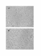
          Silicosis, caused by the inhalation of crystalline silicon dioxide or silica, is one of the most severe occupational diseases. Persistent inflammation and progressive massive pulmonary fibrosis are the most common histological changes caused by silicosis. Association of epithelial-mesenchymal transition (EMT) of hyperplastic type II epithelial cells with the fibrotic events of pulmonary fibrosis has been suggested in in vitro silica-exposed cultured cell models, patients with idiopathic pulmonary fibrosis, and bleomycin-induced experimental models. Histological features of EMT, however, are not fully described in silicotic lungs in in vivo. The purpose of this study was to demonstrate EMT of hyperplastic type II epithelial cells in the developmental process of progressive massive pulmonary fibrosis in the lungs of rats exposed to silica. F344 female rats were intratracheally instilled with 20 mg of crystalline silica (Min-U-Sil-5), followed by sacrifice at 1, 3, 6, and 12 months after instillation. Fibrosis, characterized by the formation of silicotic nodules, progressive massive fibrosis, and diffuse interstitial fibrosis, was observed in the lungs of the treated rats; the effects of fibrosis intensified in a time-dependent manner. Hyperplasia of the type II epithelial cells, observed in the massive fibrotic lesions, dominated in the lungs of rats at 6 and 12 months after the treatment. Immunohistochemistry of the serial sections of the lung tissues demonstrated positive labeling for cytokeratin, vimentin, and α-smooth muscle actin in spindle cells close to the foci of hyperplasia of type II epithelial cells. Spindle cells, which exhibited features of both epithelial cells and fibroblasts, were also demonstrated with bundles of collagen fibers in the fibrotic lesions, using electron microscopy. Increased expression of TGF-β was shown by Western blotting and immunohistochemistry in the lungs of the treated rats. These findings suggested that enhanced TGF-β expression and EMT of hyperplastic type II epithelial cells are involved in the development process of progressive massive pulmonary fibrosis during silicosis.

          Related collections

          Most cited references23

          • Record: found
          • Abstract: found
          • Article: not found

          Epithelial-Mesenchymal Transition in Cancer: Parallels Between Normal Development and Tumor Progression

          From the earliest stages of embryonic development, cells of epithelial and mesenchymal origin contribute to the structure and function of developing organs. However, these phenotypes are not always permanent, and instead, under the appropriate conditions, epithelial and mesenchymal cells convert between these two phenotypes. These processes, termed Epithelial-Mesenchymal Transition (EMT), or the reverse Mesenchymal-Epithelial Transition (MET), are required for complex body patterning and morphogenesis. In addition, epithelial plasticity and the acquisition of invasive properties without the full commitment to a mesenchymal phenotype are critical in development, particularly during branching morphogenesis in the mammary gland. Recent work in cancer has identified an analogous plasticity of cellular phenotypes whereby epithelial cancer cells acquire mesenchymal features that permit escape from the primary tumor. Because local invasion is thought to be a necessary first step in metastatic dissemination, EMT and epithelial plasticity are hypothesized to contribute to tumor progression. Similarities between developmental and oncogenic EMT have led to the identification of common contributing pathways, suggesting that the reactivation of developmental pathways in breast and other cancers contributes to tumor progression. For example, developmental EMT regulators including Snail/Slug, Twist, Six1, and Cripto, along with developmental signaling pathways including TGF-β and Wnt/β-catenin, are misexpressed in breast cancer and correlate with poor clinical outcomes. This review focuses on the parallels between epithelial plasticity/EMT in the mammary gland and other organs during development, and on a selection of developmental EMT regulators that are misexpressed specifically during breast cancer.
            Bookmark
            • Record: found
            • Abstract: found
            • Article: not found

            Silicosis.

            Silicosis is a fibrotic lung disease caused by inhalation of free crystalline silicon dioxide or silica. Occupational exposure to respirable crystalline silica dust particles occurs in many industries. Phagocytosis of crystalline silica in the lung causes lysosomal damage, activating the NALP3 inflammasome and triggering the inflammatory cascade with subsequent fibrosis. Impairment of lung function increases with disease progression, even after the patient is no longer exposed. Diagnosis of silicosis needs carefully documented records of occupational exposure and radiological features, with exclusion of other competing diagnoses. Mycobacterial diseases, airway obstruction, and lung cancer are associated with silica dust exposure. As yet, no curative treatment exists, but comprehensive management strategies help to improve quality of life and slow deterioration. Further efforts are needed for recognition and control of silica hazards, especially in developing countries. Copyright © 2012 Elsevier Ltd. All rights reserved.
              Bookmark
              • Record: found
              • Abstract: found
              • Article: found
              Is Open Access

              TGF-β1 induces human alveolar epithelial to mesenchymal cell transition (EMT)

              Background Fibroblastic foci are characteristic features in lung parenchyma of patients with idiopathic pulmonary fibrosis (IPF). They comprise aggregates of mesenchymal cells which underlie sites of unresolved epithelial injury and are associated with progression of fibrosis. However, the cellular origins of these mesenchymal phenotypes remain unclear. We examined whether the potent fibrogenic cytokine TGF-β1 could induce epithelial mesenchymal transition (EMT) in the human alveolar epithelial cell line, A549, and investigated the signaling pathway of TGF-β1-mediated EMT. Methods A549 cells were examined for evidence of EMT after treatment with TGF-β1. EMT was assessed by: morphology under phase-contrast microscopy; Western analysis of cell lysates for expression of mesenchymal phenotypic markers including fibronectin EDA (Fn-EDA), and expression of epithelial phenotypic markers including E-cadherin (E-cad). Markers of fibrogenesis, including collagens and connective tissue growth factor (CTGF) were also evaluated by measuring mRNA level using RT-PCR, and protein by immunofluorescence or Western blotting. Signaling pathways for EMT were characterized by Western analysis of cell lysates using monoclonal antibodies to detect phosphorylated Erk1/2 and Smad2 after TGF-β1 treatment in the presence or absence of MEK inhibitors. The role of Smad2 in TGF-β1-mediated EMT was investigated using siRNA. Results The data showed that TGF-β1, but not TNF-α or IL-1β, induced A549 cells with an alveolar epithelial type II cell phenotype to undergo EMT in a time-and concentration-dependent manner. The process of EMT was accompanied by morphological alteration and expression of the fibroblast phenotypic markers Fn-EDA and vimentin, concomitant with a downregulation of the epithelial phenotype marker E-cad. Furthermore, cells that had undergone EMT showed enhanced expression of markers of fibrogenesis including collagens type I and III and CTGF. MMP-2 expression was also evidenced. TGF-β1-induced EMT occurred through phosphorylation of Smad2 and was inhibited by Smad2 gene silencing; MEK inhibitors failed to attenuate either EMT-associated Smad2 phosphorylation or the observed phenotypic changes. Conclusion Our study shows that TGF-β1 induces A549 alveolar epithelial cells to undergo EMT via Smad2 activation. Our data support the concept of EMT in lung epithelial cells, and suggest the need for further studies to investigate the phenomenon.
                Bookmark

                Author and article information

                Journal
                Vet Sci
                Vet Sci
                vetsci
                Veterinary Sciences
                MDPI
                2306-7381
                30 August 2019
                September 2019
                : 6
                : 3
                : 70
                Affiliations
                [1 ]Laboratory of Pathology, School of Life and Environmental Science, Azabu University, 1-17-71 Fuchinobe, Sagamihara-shi, Kanagawa 252-5201, Japan
                [2 ]Department of Veterinary Pathology, Tottori University, 4-101 Koyama Minami, Tottori-shi, Tottori 680-8553, Japan
                [3 ]School of Nursing, University of Shizuoka, Shizuoka-shi, Shizuoka 422-8526, Japan
                [4 ]Department of Environmental Engineering, Kyoto University Graduate School of Engineering, Kyoto-shi, Kyoto 615-8530, Japan
                Author notes
                [* ]Correspondence: a-shimada@ 123456azabu-u.ac.jp ; Tel.: +81-042-754-7111
                [†]

                These authors contributed equally to this work.

                Article
                vetsci-06-00070
                10.3390/vetsci6030070
                6789520
                31480326
                ce4bcbbc-a560-48c4-88e9-a8f066ea4fe3
                © 2019 by the authors.

                Licensee MDPI, Basel, Switzerland. This article is an open access article distributed under the terms and conditions of the Creative Commons Attribution (CC BY) license ( http://creativecommons.org/licenses/by/4.0/).

                History
                : 06 August 2019
                : 27 August 2019
                Categories
                Article

                epithelial-mesenchymal transition,fibrosis,rat,silicosis,tgf-β,type ii epithelial cells

                Comments

                Comment on this article